
Indications
- excision of herniated discs
- exploration of nerve roots
- spinal fusion
- removal of tumours
Position
- prone
- bolsters longitudinally under patient’s sides
- abdomen free
- reduces venous plexus filling around the spinal cord by permitting the venous plexus to drain directly into the vena cava
- abdomen free
- Mayfield head rest
- GA
Landmarks
- Spinous processes
- Iliac crest = L4-5 interspace
- Identify Level with II
Incision
- Midline longitudinal incision
- Start: depends on numbers of levels to be explored
Internervous Plane
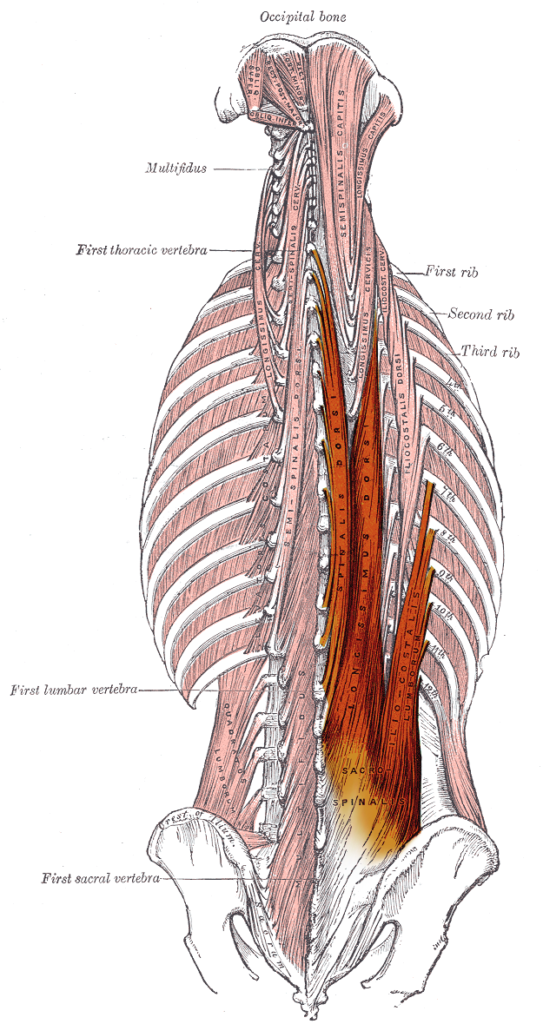
- Between 2 paraspinal muscles (erector spinae)
- Receive segmental nerve supply from posterior primary rami of lumbar nerves
Superficial Dissection
- Inline with incision
- Fat
- Fascia
- Reach spinous process
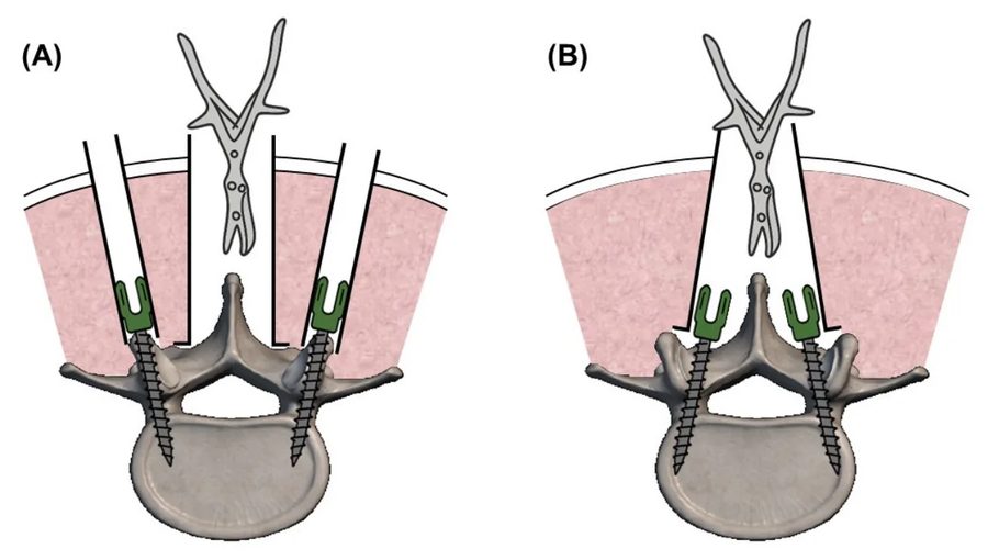
- Detach paraspinal muscles subperiosteally using Cobb elevator
- Dissect down the spinous process, lamina, facet joint
- If needed
- Strip facet joint capsule
- Transverse process
Deep Dissection
- Ligamentum flavum
- Cutting attachment to the superior edge of the inferior lamina
- Beneath it is
- Epidural fat
- Blue-white dura
- Remove the rest of the ligamentum flavum & undersurface of lamina with a Kerrison Leskel
- Stay lateral to dura
- Identify nerve root
Dangers
- Segmental vessels supplying paraspinal muscles
- Posterior primary rami of lumbar nerves
- Nerve roots
- Venous plexus
- Stop with gelfoam, cotton patties soaked in thrombin
- Bipolar malis cautery
- Iliac vessels
- Injured if instruments pass through anterior portion of annulus fibrosus
- Local measures
- Remove additional portions of lamina
- Remove portion of facet joint
- Carry dissection far lateral onto transverse process
- Extensile
- From C1 to sacrum
Reviewed by Professor Phong Tran, Head of Orthopaedic Surgery, Western Health. Last updated 10 October 2026.