
Indications
- Fusing L5 to S1
- Not for occasional user
Position
- supine
- prep & drape for 2 incisions
- abdominal
- anterior iliac crest bone graft
- GA
- urinary catheter
- nasogastric tube
- ileus is common
Landmarks
- umbilicus
- opposite L3-4 disc space
- pubic symphysis
Incision
- longitudinal midline incision
- Start: xiphisternum
- Curved to the LEFT around umbilicus
- End: pubic symphysis
Internervous Plane
- Abdominal muscles on each side
- Segmentally supplied by branches from 7th to 12th intercostal nerves
Superficial Dissection
- Deepen wound in line with incision
- Fat
- Rectus sheath
- Incise longitudinally
- Reveals 2 rectus muscles
- Separate with fingers
- Peritoneum
- Pick up with pair of forceps & incise with knife
- Extend incision watching for viscera, bladder
Deep Dissection
- Balfour self retaining retractor
- Retract viscera laterally
- Put table in Trendelenburg’s position of 30°
- Carefully pack bowel cephalad
- Infiltrate tissue over anterior surface of sacral promontory with a few millilitres of saline
- Makes dissection easier
- Helps identify presacral parasympathetic nerves
- Ligate sacral artery
- Runs down midline
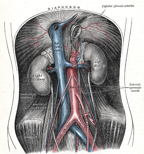
- Identify L5-S1 disc space
- Palpate sharp angle
- I.I
- Lies below aorta bifurcation
Dangers
- Presacral plexus of parasympathetic nerves
- Retroejaculation & impotence
- Vessels
- Middle sacral artery
- Aorta
- IVC
- Ureter
Reviewed by Professor Phong Tran, Head of Orthopaedic Surgery, Western Health. Last updated 10 October 2026.