
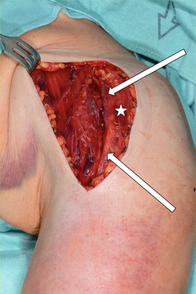
The anterior (deltopectoral) approach to the shoulder uses the internervous plane between deltoid, supplied by the axillary nerve, and pectoralis major, supplied by the pectoral nerves. It is the workhorse approach for shoulder arthroplasty, proximal humerus fixation and anterior stabilisation, protecting the cephalic vein and musculocutaneous nerve.
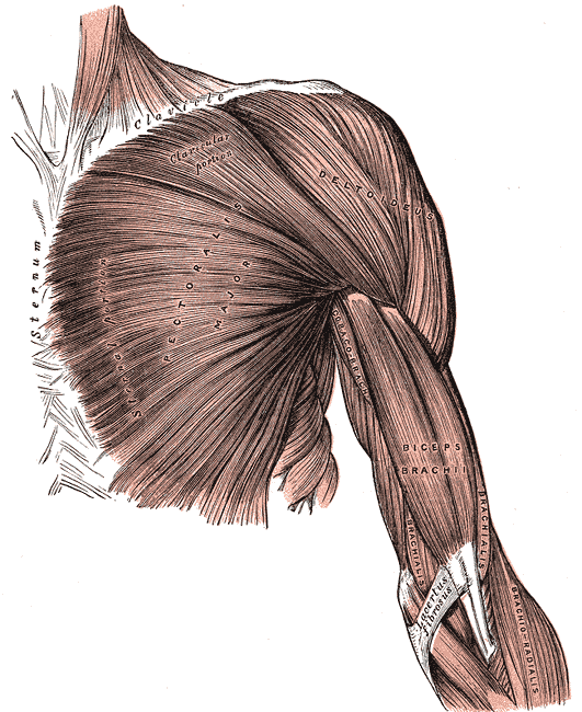
Position
- supine
- sandbag under spine & medial border of scapula
- beachchair to 30°
- Mayfield head rest
- Prep & drape arm free
Landmarks
- Clavicle
- Acromion
- Coracoid process
- Deltopectoral groove
Incision
1. Anterior incision
- Start: just above coracoid process
- 10-15 cm along line of deltopectoral groove
2. Axillary incision
- abduct shoulder 90° & ER
- start: midpoint of anterior axillary skin fold
- vertical incision 8-10 cm
- end: towards axilla
Internervous Plane
- deltoid: axillary nerve
- pectoralis major: medial & lateral pectoral nerves
Superficial Dissection
- identify deltopectoral groove
- cephalic vein laterally
- deltoid laterally
- pect major medially
Deep Dissection
- conjoint tendon: short head of biceps (MC nerve), coracobrachialis (MC nerve) taken medially
- Dangers:
- Axillary artery lies behind pect minor
- Keep arm adducted during procedure to protect artery
- Musculocutaneous nerve
- On medial side of coracobrachialis
- Axillary artery lies behind pect minor
- Deltopectoral fascia
- Subscapularis tendon
- ER arm to stretch subscapularis & make it easier to define
- Also ↑ gap between subscapularis & axillary nerve (at its lower border, quadrangular space)
- Ligate triad of small vessels*2 veins 1 artery) at inferior border
- Superior border of SubS is indistinct & blends with supraspinatus
- Tag tendon
- Detach subscapularis off lesser tuberosity insertion
- Capsule
- Longitudinal incision
Dangers
- Musculocutaneous nerve
- Enters CB 5-8 cm distal to coracoid process
- From medial side
- Cephalic vein
Reviewed by Professor Phong Tran, Head of Orthopaedic Surgery, Western Health. Last updated 10 October 2026.