
Indications
- excellent exposure of
- talocalcaneonavicular
- posterior talocalcaneal
- calcaneocuboid joints
- arthrodesis
- including triple arthrodesis
Position
- supine
- sandbag
- exsanguination
- tourniquet
Landmarks
- lateral malleolus
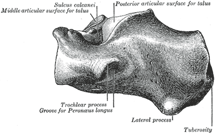
- lateral wall of calcaneus
- sinus tarsi
Incision
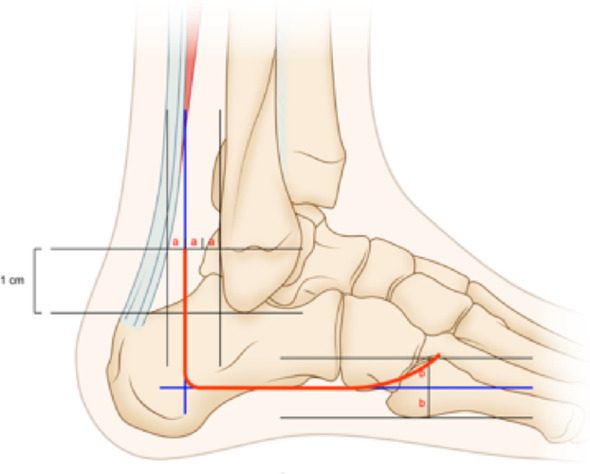
- curved incision
- Start: just distal to distal end of lateral malleolus
- Over sinus tarsi, curving medially
- End: talocalcaneonavicular
Internervous Plane
- Peroneus tertius
- Peroneal tendons
Superficial Dissection
- Do not mobilise skin flaps
- Ligate veins
- Open deep fascia in line with skin incision
- Beware
- Peroneus tertius
- EDL
- Retract these muscles medially
- Beware
Deep Dissection
- Detach fat pad from sinus tarsi
- Detach EDB from calcaneus
- Expose capsule of talocalcaneonavicular & calcaneocuboid joint
- Open these joints as necessary
- Incise peroneal retinacula
- Reflect peroneal tendons anteriorly
- Incise capsule of posterior talocalcaneal joint
Dangers
- Skin flap NECROSIS ++
Extensile
- Proximally
- Curve along the posterior border of fibula
- Develop plane between peroneal muscles & flexor muscles
Reviewed by Professor Phong Tran, Head of Orthopaedic Surgery, Western Health. Last updated 10 October 2026.