
Aims
- Exposure of the lateral malleolus
Indications
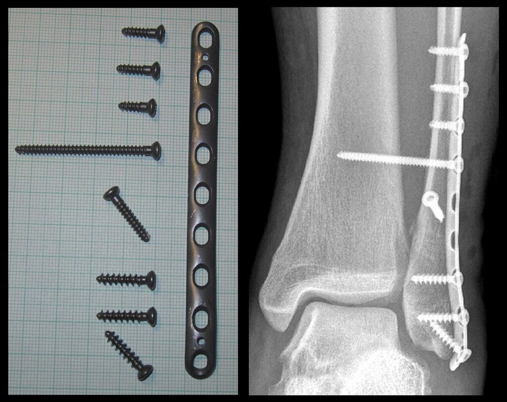
- Fracture Fixation
Position
- supine
- sandbag under buttock
- exsanguinate
- tourniquet
Landmarks
- subcutaneous surface of fibula
- lateral malleolus
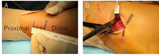
Incision
- 10-15cm incision along posterior margin of fibula
Internervous Plane
- None
- More proximally
- Peroneus tertius deep peroneal nerve
- Peroneus brevis superficial peroneal nerve
Superficial Dissection
- Elevate skin flaps
- Beware
- Short saphenous vein
- Sural nerve
- Beware
Deep Dissection
- Incise periosteum
- Beware
- Terminal branches of peroneal artery
Dangers
- Sural nerve
- Terminal branches of peroneal artery
Procedure
- Proximal
- Posterior border of fibula
- Distal
- Curve incision over lateral side of foot
- Identify peroneal tendons & incise peroneal retinaculum
- Detach fat pad from sinus tarsi
- Detach origin of EDB to expose CC joint
- Curve incision over lateral side of foot
Reviewed by Professor Phong Tran, Head of Orthopaedic Surgery, Western Health. Last updated 10 October 2026.