
Classification
| Tumour | Features |
|---|---|
| Lipoma | Most common soft tissue tumour, superficial, soft and mobile |
| Intramuscular lipoma | Deep, may infiltrate muscle |
| Atypical lipomatous tumour | Deep and large, MDM2 amplification, local recurrence without metastasis |
| Myxoid liposarcoma | Metastasises to unusual sites such as spine and retroperitoneum. Whole spine MRI for staging |
| Dedifferentiated and pleomorphic liposarcoma | High grade, metastatic potential |
Clinical Features
- Painless, slowly enlarging mass in most cases
- Lipomas are common on the back, shoulders and thigh
- Liposarcoma is most common in the thigh and retroperitoneum in middle aged adults
Red Flags
- Size over 5 cm
- Deep to fascia
- Increasing size
- Pain
- Refer to a sarcoma service before biopsy
- Any soft tissue mass meeting these features needs MRI before excision
- Unplanned excision of a sarcoma compromises limb salvage
Investigations
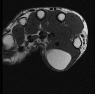
- MRI, simple lipomas follow fat signal and suppress fully
- Thick septa, nodules or enhancement suggest liposarcoma
- Ultrasound for small superficial lesions
- MDM2 amplification on FISH distinguishes atypical lipomatous tumour from lipoma
- Core biopsy along the line of future excision at a sarcoma centre
- CT chest for staging of liposarcoma
Management
- Lipoma, marginal excision if symptomatic
- Atypical lipomatous tumour, marginal excision
- Liposarcoma, wide excision with radiotherapy
- Atypical lipomatous tumour in the retroperitoneum behaves more aggressively than in the limbs
- Myxoid liposarcoma is radiosensitive, and preoperative radiotherapy is common
- Follow up with clinical review and imaging for recurrence
Related Pages
Reviewed by Professor Phong Tran, Head of Orthopaedic Surgery, Western Health. Last updated 10 October 2026.