
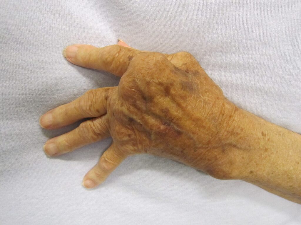
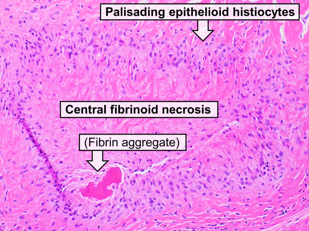
Definition
- Chronic symmetrical polyarthritis of unknown origin
Epidemiology
- Worldwide distribution & affects all races & ethnic groups
- Most common inflammatory arthropathy
- 3% of population
- M:F – 1:3
- Annual incidence of 2-4/ 10 000 adult population
- Peak age 4th-6th decade
- Peak onset 40 years
- Increasing in West
- Life span ↓ 3-18 years
Aetiology
- Unknown cause
- Cell mediated immune response
- Postulated combination of genetic susceptibility & environmental trigger
- External trigger
- ? Infection
- Viral aetiology suggested due to synovitis similar to RA in patients with
- Hepatitis B
- Ross river virus
- Rubella
- Abnormal immune response in RA patients to EBV
- Mycobacteria & Lyme disease related but no bacteria isolated from the joints in septic arthritis
- Can induce arthritis in rats with immunisation with
- Streptococcal cell walls
- Soluble type II collagen
- Freunds adjuvant
- Genetic susceptibility
- Associated with HLA DR4
- 70% Rheumatoid Arthritis
- 30% Normal
- 80% of patients with RA are HLA DW4 and/ or D4 positive
- Concordance amongst identical twins is 30%
- Some large series suggest incidence of RA in first degree relatives twice that of chance alone
- Associated with HLA DR4
- Exogenous agent alters IgG to become antigenic
- Helper T cells activate B Cells to become plasma cells
- Plasma Cells produce Rheumatoid Factors (IgM usually) directed against IgG
- Synovium acts as lymphoid organ
- Local helper T cells activated
- Local plasma cells produce RF
- Antibody-Antiglobulin complexes formed
- Stimulates complement
- Secondary destructive inflammatory cascade
- Rheumatoid Factor
- Antiglobulin directed at Fc region of IgG
- Production of antigens on surface of IgG suggested to be due to infective or other trigger
- Usually IgM but heterogeneous
- Can be IgM, IgG, IgA or IgE
- Seen in 80% with Rheumatoid Arthritis
- Incidence of RF positivity ↑ with duration of active synovitis
- Correlation with
- Long standing disease
- Sustained activity
- Nodules
- Erosions
- Extraarticular manifestations
- ? Role in pathogenesis
- Synthesised locally in subsynovial tissue & demonstrable in plasma cell by immunofluorescence & as eosinophilic bodies on H & E (Russell Bodies)
- Synovium thus acts as lymphoid tissue like spleen & lymph nodes
- Detected with Latex Agglutination
- Use latex particles with human IgG adsorbed to surface
- Various dilutions of test sera to agglutinate the complexes
- Titre is highest dilution that agglutinates
- 1:80 is positive
- High Titre » Severe Disease
- May also be present in
- Elderly (incidental)
- Infection
- Autoimmune Disease
- Chronic liver disease
- Multiple Myeloma & other Lymphoproliferative disorders (5-10%)
Reviewed by Professor Phong Tran, Head of Orthopaedic Surgery, Western Health. Last updated 10 October 2026.