
Base of thumb injuries include Bennett and Rolando fractures of the first metacarpal base, extra-articular base fractures and carpometacarpal dislocations. The thumb provides up to 40% of hand function, so intra-articular fractures are reduced accurately, usually with K wires or screw fixation.
Background
- Thumb provides up to 40% of hand function
- Total disability = loss of 22% of bodily function
- CMC is biconcave-convex saddle-shaped joint
- Axis of MC is pronated and flexed 80o relative to other MC
Imaging
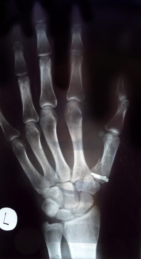
- True AP – Robert’s view
- True lateral – Bett’s view
- Oblique view
- Kapandji PA view
Fractures
- Most common in young (0-16) and elderly (65+)
- Classification:
- Extra-articular
- Partial intra-articular (Bennett)
- Complete intra-articular (Rolando)
- Severely comminuted fracture
Extra-articular
- Metadiaphyseal junction fractures most common (epibasal fracture)
- Apex dorsal angulation due to AddP, FPL & APB on distal fragment
- Accept up to 30o angulation
- Anything more = compensatory MCP hyperextension
- Management:
- Closed reduction – axial traction, extension & pronation with direct pressure over fracture
- ORIF – k-wires, lag screw, plate, ex-fix
Bennett
- Described by E.H. Bennett in 1882
- Intra-articular 2 part fracture – volar ulnar fragment
- Fragment held by anterior oblique ligament attachment to trapezium (beak ligament)
- MC shaft subluxes dorsal/proximal/radial
- Mechanism:
- Axial load on partially flexed MC
- Look for trapezium fractures and UCL injuries
Gedda Classification
| Type | Description |
|---|---|
| 1 | Large single ulnar fragment with subluxation of MC base |
| 2 | Impaction fracture without subluxation of MC base |
| 3 | Small ulnar avulsion fragment with CMC dislocation |
Management
- Closed reduction – axial traction, abduction and pronation with pressure over MC base
- Tensions dorsal ligament complex to reduce (Edmunds, 2006)
- Poorer outcomes with casting alone (Kjaer-Peterson et al. 1990)
- Closed reduction + intermetacarpal fixation to 2nd MC and/or trapezium
- Can add k-wire through volar ulnar fragment
- Open reduction if >1-2mm displacement/intra-articular step (controversial in literature)
- Wagner approach
- Consider distraction + ex-fix for fragments too small to fix
Rolando
- Described by Silvio Rolando in 1910
- Y or T shaped 3 part intra-articular fracture
- Volar ulnar fragment + dorsal radial fragment
- Worse prognosis – over 50% CMC OA (Langhoff et al. 1991)
- Management:
- CRIF for simple 3 part fractures if <1mm displacement
- ORIF if >1mm displacement
- Distraction + fixation/ligamentous reduction for highly comminuted fractures
- Traction pinning, external fixation
1st CMC Dislocation
- Rare (<1% of thumb injuries)
- Mostly dorsal
- Mechanism:
- Axial force on flexed thumb
- Dorsal force through 1st web space (e.g. handlebar into thumb)
- Presentation:
- Pain, swelling and bruising over thenar eminence
- Unable to form fist
Anatomy
- 16 ligaments stabilise CMC joint — 4 important ones:
- Dorsoradial ligament check rein to radial subluxation (most important)
- Anterior oblique ligament (superficial/deep)
- Posterior oblique ligament
- Intermetacarpal ligament
Investigations
- Standard radiographs
- MRI
- Persistent/recurrent instability post reduction
- Guides ligament reconstruction
Management
- Closed reduction + immobilisation in extension/pronation
- Only if stable on reduction
- Closed reduction + percutaneous pinning
- Recommended treatment
- For more unstable injuries
- Adding dorsal capsuloligamentous reconstruction with FCR autograft + pinning leads to better strength & ROM and lower pain (Simonian & Trumble 1996)
- Low incidence of recurrent dislocation
Ulnar Collateral Ligament Injuries
- Thumb MCP joint stable throughout flexion/extension arc
- Range of motion is extremely variable – even between sides in same pt
- Consists of two parts:
- Proper collateral ligament – taut in flexion
- Accessory collateral ligament – taut in extension
- Dynamic stability from adductor pollicis, FPB and EPB
- Mechanism of injury
- Excessive radial deviation at MCP joint
- Acute injury = skier’s thumb
- Usually distal avulsion
- 50% have P1 fractures
- Chronic injury = gamekeeper’s thumb
Physical Exam
- Can differentiate complete from partial tears
- Difficult in acute injuries due to pain/spasm
- Unstable injury:
- >35o joint angulation on valgus stress of flexed MCP = complete proper collateral lig tear
- >35o joint angulation on valgus stress of extended MCP = complete accessory collateral lig tear
- Greater than 20o variation in side to side valgus laxity
- Lack of firm end point on stress testing
Stener Lesion
- UCL torn and displaced proximal/superficial to adductor pollicis aponeurosis
- Aponeurosis interposed between ligament and attachment point = unable to heal
- Diagnosis:
- Palpable mass proximal to MCPJ
- XR – may see bony Stener lesion
- MRI – specificity 95%, sensitivity 96%
- Indication for surgical repair
Treatment
- Acute:
- Stable injuries/partial tears – 4 weeks immobilisation spica splint/cast
- Unstable injuries/complete tears – surgical repair
- Suture repair of ends vs suture anchor repair of avulsions
- Avulsion fractures with no bony Stener lesion = controversial
- Generally non-op for undisplaced fractures that are stable on stress testing
- Be careful with stress testing – may turn undisplaced into displaced lesion
- Consider leaving for 1-2 weeks then stress testing once pain settled and lesion not as mobile
- Variable outcomes, painless non-unions, ongoing instability
- Generally non-op for undisplaced fractures that are stable on stress testing
- Chronic (gamekeeper’s thumb)
- Reconstruct only in absence of significant MCPJ arthritis
- Up to 2yrs can consider mobilisation of UCL from scar and repair to bone with anchors
- Dynamic procedures – utilising adductor pollicis or EPB
- Static procedures – free tendon grafts to reconstruct ligaments
Complications
- General:
- Reduced grip/pinch strength and reduced function
- Post-traumatic OA and pain/stiffness
- Fractures
- Malunion/non-union
- Deformity
- CMC dislocation + UCL injury
- Recurrent instability
- Surgical
- Injury to dorsal branches of SRN
- Pin site infection
- Failed repair/reconstruction
References
- Carlsen BT & Moran SL. Thumb trauma: Bennett fractures, Rolando fractures, and ulnar collateral ligament injuries. J Hand Surg Am. 2009 May-Jun;34(5):945-52.
- Edmunds JO. Traumatic dislocations and instability of the trapeziometacarpal joint of the thumb. Hand Clin. 2006 Aug;22(3):365-92.
- Kjaer-Peterson K, Langhoff O, Andersen K. Bennett’s fracture. J Hand Surg Br. 1990 Feb;15(1):58-61.
- Langhoff O, Andersen K, Kjaer-Peterson K. Rolando’s fracture. J Hand Surg Br. 1991 Nov;16(4):454-9.
- Simonian PT & Trumble TE. Traumatic dislocation of the thumb carpometacarpal joint: early ligamentous reconstruction versus closed reduction and pinning. J Hand Surg Am. 1996 Sep;21(5):802-6.
Contributions
Page written by Dr James Drummond (orthopaedic registrar) 2020
Reviewed by Professor Phong Tran, Head of Orthopaedic Surgery, Western Health. Last updated 10 October 2026.