
Definition
- Juvenile rigid kyphosis
- Structural thoracic or thoracolumbar kyphosis
- Endplate growth disturbance causes anterior vertebral wedging
Epidemiology
- Presents in adolescence, around 10 to 15 years
- One of the most common causes of structural kyphosis at this age
- Male predominance in many series
- Familial tendency
Aetiology
- Cause unknown
- Proposed factors are genetic predisposition, mechanical loading, endplate osteoporosis and disordered ossification of the ring apophysis
Classification
- Typical (thoracic), apex mid thoracic
- Atypical (thoracolumbar or lumbar), more often painful, associated with heavy physical activity
Clinical Features
- Round back deformity, often noticed by parents
- Aching thoracic pain worse with activity
- Rigid kyphosis not correcting on prone hyperextension, distinguishing it from postural kyphosis
- Sharp angular deformity on forward bending
- Compensatory lumbar and cervical hyperlordosis
- Hamstring tightness and increased rate of spondylolysis
- Neurology rare, from thoracic disc herniation or epidural cyst
Investigations
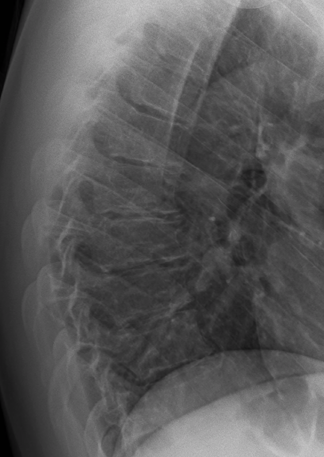
- Sorensen criteria, anterior wedging of 5° or more in at least three adjacent vertebrae
- Thoracic kyphosis above about 45°
- Schmorl nodes, endplate irregularity and disc space narrowing
- Mild scoliosis in some
- Hyperextension lateral over a bolster assesses flexibility
- MRI for neurological deficit and before surgery
Management
- Observation and physiotherapy for mild curves and pain
- Brace skeletally immature curves of about 50° to 75° in a Milwaukee brace or modified TLSO until maturity
- Bracing is less effective for rigid or large curves and after maturity
- Surgery for thoracic kyphosis over about 75° or thoracolumbar over about 65° with progression, pain, deformity or neurology
- Posterior fusion with segmental pedicle screws and Ponte osteotomies
- Add anterior release for very large rigid curves
- Fuse from the proximal end vertebra to the sagittal stable vertebra to reduce junctional kyphosis
- Avoid overcorrection, which increases proximal junctional kyphosis
Reviewed by Professor Phong Tran, Head of Orthopaedic Surgery, Western Health. Last updated 10 October 2026.