
Definition
Fibroproliferative disorder of palmar aponeurosis
Aetiology
Murrell’s Theory of Pathogenesis
- Initiating event is microvascular ischaemia
- Ischaemia itself leads to conversion of
- ATP toHypoxanthine
- Endothelial Xanthine Hydrogenase to Xanthine Oxidase
- Xanthine Oxidase catalyses reaction
- Hypoxanthine to Uric Acid
- Which gives off OH- (hydroxyl free radicals)
- OH- stimulate fibroblast proliferation
- Increasing Type III Collagen
- Fibroblasts strangle microvessels
- Vicious Cycle
- ATP: Hypoxanthine: Uric acid + OH-: fibroblast proliferation: microvascular strangulation
Epidemiology
- 5% Caucasians
- Especially Vikings
- Rare Blacks & Asians
- M:F – 5:1
- 20% Male > 65 years
Risk Factors
- Local ischaemia
- DM
- Smoking
- Trauma (including fractures of the wrist)
- Occupation? (weight of evidence against it)
- Alcoholic cirrhosis
- Anti-epileptic medication (phenobarbitone)
- HIV
- Genetic
- Vikings – Anglo-Saxon/ Celtic origin
- AD with variable penetrance
Anatomy
- Key to dissection
- Normal structures (bands) become cords & nodules
- Mnemonic “bland bands become crazy cords”
Pretendinous Bands
- Volar & midline to neurovascular bundle
- Form from palmar aponeurosis as it travels to digits
- Splits at MCPJ
- Some into skin
- Rest deep to neurovascular bundle as spiral band
Natatory Ligament
- Transverse & superficial to neurovascular bundle at web space
- Attached to flexor sheath/ skin
- Almost always involved
Spiral Bands
- Termination of pretendinous band
- Pass deep then lateral to neurovascular bundle
Lateral Digital Sheet
- Lateral condensation superficial fascia
Grayson’s Ligament
- Volar to neurovascular bundle
- Arises tendon sheath
- Inserts to lateral digital sheet
Cleland’s Ligament
- Dorsal to nv structures & same origins as Grayson’s ligaments
- Not involved
Transverse Fibres of the Palmar Aponeurosis
- “Skoogs fibres”
- Superficial to nv bundles at level MC heads
- Never involved
- Where you should look to trace nv bundles
Cords
- Central, Spiral, Lateral, Natatory
- Form along normal fascial pathways
- Tighten up
- Follows three dimensional anatomy
- Neurovascular bundle spirals around spiral cord
- Between distal palm crease & proximal crease finger
- Coalesce into one
Spiral Cord
- Formed by
- Pretendinous band (medial to nv bundle)
- Spiral Band (deep)
- Lateral Digital Sheet (lateral)
- Grayson’s ligament (superficial)
Pathology
Myofibroblasts
- Contractile cell
- Occur elsewhere (GUT)
- But not normally in palmar fascia
- Originate as Perivascular Fibroblasts
- Convert secondary to local hypoxia
- Mainly in nodules
- None in cords
- Responds to cell growth factors
- PDGF/ FGF/ TGF B
Type III Collagen
- Increased secondary to ↑ cell density
- Controversial role
Abundant Fibroblasts
- Cords only
- Peri-arterial
- Organised along stress lines
- Occluded microvessels
Classification
Tubiana, by total flexion deformity of MCP, PIP and DIP joints
| Stage | Total flexion deformity |
|---|---|
| N | Nodule without contracture |
| 1 | 0 to 45 degrees |
| 2 | 45 to 90 degrees |
| 3 | 90 to 135 degrees |
| 4 | Over 135 degrees |
Original publication Tubiana R. Evaluation of deformities in Dupuytren’s disease. Ann Chir Main. 1986;5(1):5-11.
History
- M>F – 7:1
- Right > Left
- RF > LF > MF > thumb > IF
- Tender nodules beyond distal palmar crease
- Painless fixed flexion deformity with disability
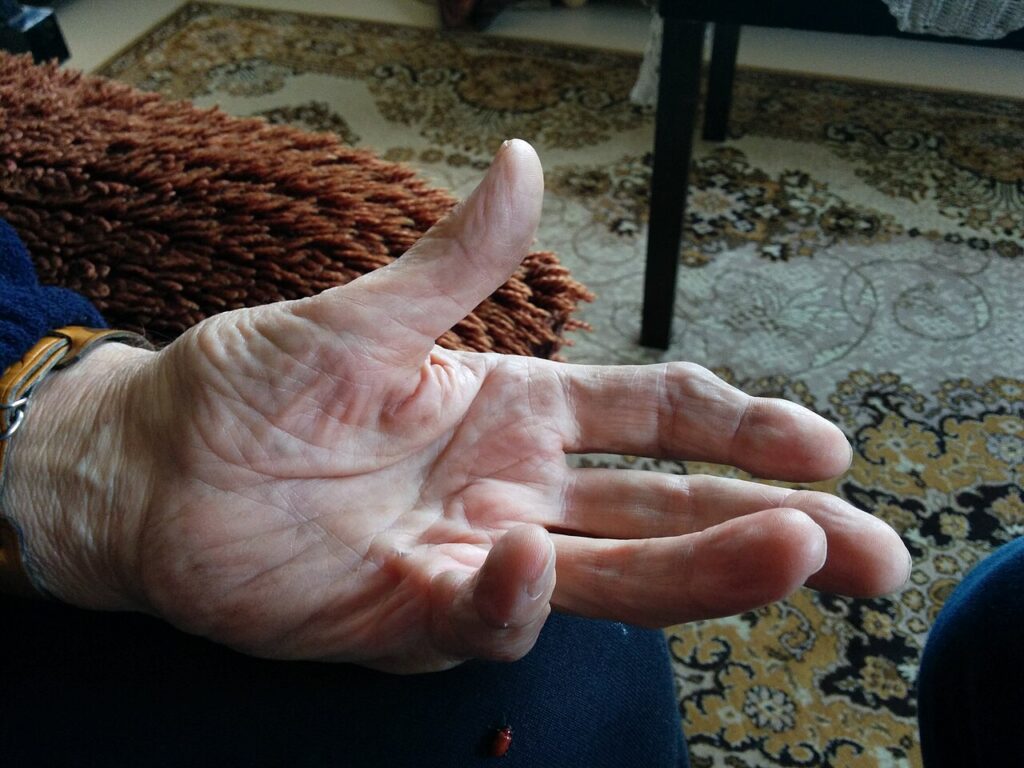
- Nodules, cords, pits
Dupuytren’s Diathesis
- Aggressive Disease: rapid progression & marked contractures
- Young males
- Significant family history
- Multiple digits
- Bilateral
- Garrod’s Nodes (dorsal thickening of knuckle pads at PIPJ vs carpet layers at MCPJ)
- Lederhose Disease
- Peyronie’s Disease
Examination
- Palpable nodules and cords, ring and little fingers most often involved
- Goniometer measurement of MCP and PIP flexion deformity
- Hueston table top test positive when the palm cannot lie flat
- Skin pitting and blanching on finger extension
- Ectopic disease with Garrod knuckle pads, Ledderhose plantar fibromatosis and Peyronie disease
- Digital sensation and perfusion before surgery, especially in recurrent disease
- Thumb and first web space involvement
Investigations
- Clinical diagnosis, imaging not required
- US can confirm a nodule when the diagnosis is uncertain
- Blood glucose and liver function tests when diabetes or alcohol excess suspected
- X-ray when PIP joint arthritis is suspected
Differential Diagnosis
- Trigger finger
- Camptodactyly
- Post traumatic PIP joint contracture or boutonnière deformity
- Ulnar nerve palsy clawing
- Ganglion, epidermoid cyst or giant cell tumour of tendon sheath
- Epithelioid sarcoma
- Palmar callus
Treatment
Don’t Operate If Negative Table Top Test
- Don’t operate if nodule is painful – can trigger surrounding tissue & ↑ proliferation
- On occasion may need to operate if deep skin folds & pits cause repeated maceration & infection
Non-operative
- No proven benefit
- Corticosteroids (to nodules & Garrod’s pads – Ketchum 1991)
- Occupational Therapy
- Allopurinol
- 5-FU
- Vitamin E
- Patient Education
Operative
Hueston’s “Table Top” Test
- Palm unable to touch table when trying to place hand flat on table
- If FFD of PIPJ & MCPJ able to hyperextend & allows palm to touch table then this is a –ve test
- Positive when MCPJ fixed flexion deformity > 40°?
MCPJ fixed flexion deformity
- easily correctable (see below)
- Often becomes troublesome with FFD > 30°
- Generally correctable at any degree of FFD
PIPJ fixed flexion deformity
- difficult to correct
- 30° fixed flexion deformity usual end result
- Hence McFarlane (1990) advocates
- Release of PIPJ only if fixed flexion deformity > 30°
- Preoperative splint?
Three Technical Considerations
- Incision
- Fasciotomy/ fasciectomy
- Fasciotomy
- Segmental fasciectomy
- Selective fasciectomy
- Radical fasciectomy
- Wound closure
“Operation is dissection of the neurovascular bundles”
Skin Options
- Direct Closure ± Flaps
- Longitudinal with Z-plasty
- Midline longitudinal
- Points at creases in midlateral line passing to midline
- Angle 60° to longitudinal axis
- Allows dissection from palm to finger
- Does not allow wide dissection in palm: ↓ postoperative haematoma
- Transverse
- Used in palm when 2-3 rays involved
- Can be combined with longitudinal incisions
- Simple, good exposure & rapid healing
- Skin necrosis & ↑ postop haematoma
- Brunner ± VY plasty
- Popular
- Can do lazy Brunner (lazy “S”)
- Zig-zag prevents scar contracture
- However difficult to design flaps with finger contracted & flap difficult to construct with intimate proximity of diseased fascia
- Longitudinal with Z-plasty
- Dermofasciectomy
- Skin excision
- Full thickness flaps
- Less recurrence
- Graft failure
- McCash Open Technique
- Transverse mid palm wound at distal palmar crease
- Leave open but review regularly
- Allows haematoma to drain
- Closes by contraction (takes 3-5/52)
Fascia Options
- Fasciotomy/ Segmental Fasciectomy
- Limited procedure with cord divided or short portion of fascia excised
- Partial or complete correction
- Elderly or mild disease
- Can be performed as outpatient procedure
- Selective Fasciectomy
- Only Dupuytren’s tissue excised – leave normal looking tissue
- Most popular technique
- Mainstay
- Subclinical disease may progress but often does not warrant further surgery
- Radical Fasciectomy
- Excision of normal & diseased fascia
- Less popular now
- Still get recurrence
- For those with Dupuytren’s diasthesis?
Wound Closure
- Options
- Suture
- Graft
- Leave open
- Principle is closure without tension as this predisposes haematoma & wound breakdown
Release of PIPJ FFD
- Volar capsule/ volar plate released proximally at pars flaccida
- Checkrein Ligament (at proximal end of volar plate) released
- Accessory Collateral Ligaments (“sides of the wheelbarrow”) released
- Then pass along volar portion of collateral ligaments (ie “remove the arms off the wheelbarrow”)
- Then can release one collateral ligament if still tight
Management of Recurrence
- Revision
- Allen’s Test Finger
- Test Sensation
- High risk neurovascular bundle injury
- Selective fasciectomy & graft
- PIPJ fusion if severe
- Amputation of finger if
- PIPJ fixed flexion deformity > 90°
- Insensate
- Pain
- Dysvascular
Complications
- 20%
- Classic triad
- Haematoma
- Infection
- Skin loss
- Neurovascular bundle injury
- Start dissection of nerve proximally
- Recurrence 50%
- Dupuytren’s in a surgically treated field
- 15% need repeat operation
- RSD
Prognosis
- Progressive in most, at an unpredictable rate
- Recurrence at 5 years about 85% after needle fasciotomy and 21% after limited fasciectomy (van Rijssen)
- Recurrence at 5 years about 47% after collagenase (CORDLESS)
- MCP contractures correct more completely than PIP contractures
- Dupuytren diathesis predicts early recurrence and extension
Reviewed by Professor Phong Tran, Head of Orthopaedic Surgery, Western Health. Last updated 10 October 2026.