
Definition
- Fracture of the Sacrum
Incidence
- Frequent component of pelvic ring injuries
- Isolated sacral fractures are less common
- Bimodal distribution with young high energy and elderly insufficiency fractures
- Insufficiency fractures are rising with an ageing population
Aetiology
- High energy trauma such as motor vehicle accidents and falls from height
- Insufficiency fractures with osteoporosis, pelvic radiotherapy and corticosteroid use
- Stress fractures in athletes and military recruits
- Fall from height onto the buttocks causes transverse and U shaped fractures
Anatomy
- Five fused sacral vertebrae articulating with the ilia at the SI joints
- Upper sacral bodies accept iliosacral screws within a safe corridor
- Sacral foramina transmit the ventral rami of the sacral nerves
- L5 nerve root crosses the anterior sacral ala
- Upper sacral dysmorphism narrows the safe screw corridor
- Posterior SI ligaments are the strongest ligaments of the pelvic ring
Classification
- Vertical, oblique or transverse
- Vertical
- Denis classification of Vertical fractures
- 3 Zones
- Denis classification of Vertical fractures
- Vertical
| Zone | Description | Nerve Injury |
|---|---|---|
| 1 | lateral to neural foramen | 5% rate of nerve injury (L5 usually) |
| 2 | through neural foramen | 25% rate of nerve injury |
| 3 | medial to foramen | 50% rate of nerve injury – involves central sacral canal |
- Transverse fractures
- Usually S12
- High rate of neurological injury
- If S23 may lead to loss of bladder and bowel function
- Pelvic and lower lumbar spine injuries often present
- Usually S12
Pathology
- Vertical fractures form part of a posterior pelvic ring disruption
- Transverse and U shaped fractures cause spinopelvic dissociation
- Nerve injury from foraminal or canal compromise
- Neurological deficit increases from zone I to zone III
- Bilateral S2 to S4 injury causes bladder, bowel and sexual dysfunction
History
- Mechanism of Injury
- Sensory disturbance
- Bowel | bladder function
Examination
- Neurological Examination
- PR
- Identify associated injuries
Investigations
X-rays
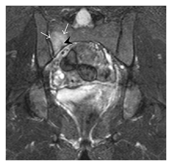
- often difficult to see
CT Scan
- bone anatomy
MRI
- soft tissue anatomy
Treatment
Non-operative
- Stable fractures, bed rest
- X-ray monitoring
Operative
- Unstable or displaced
- CR & percutaneous SI screws
- Trans iliac bars
- Plate and screws
- +/- laminectomy
Complications
- Persistent neurological deficit with bladder, bowel and sexual dysfunction
- Malunion with leg length discrepancy and pelvic obliquity
- Non union
- Screw malposition with L5 or S1 root injury
- Wound breakdown after posterior approaches, especially with Morel Lavallée lesions
- VTE
Prognosis
- Stable isolated fractures heal with good function
- Neurological injury predicts a poorer outcome
- Displaced vertical fractures and spinopelvic dissociation often leave pain and gait disturbance
- Most insufficiency fractures heal with mobilisation
- Elderly insufficiency fractures carry raised mortality
Reviewed by Professor Phong Tran, Head of Orthopaedic Surgery, Western Health. Last updated 10 October 2026.