
Pelvic ring injuries range from stable low energy fractures in older people to high energy disruptions with major haemorrhage. They are classified by mechanism with Young and Burgess or by stability with Tile. Unstable injuries need resuscitation, a pelvic binder and external or internal fixation.
Definition
Western Health Orthopaedic Registrar presentation – Fractures of the Pelvis and Acetabulum by Dr Peter Moore
Incidence
- About 3% of skeletal injuries
- Bimodal, young patients with high energy trauma and elderly patients with fragility fractures
- Associated injuries common after high energy mechanisms
Aetiology
- Pelvic ring injuries are caused by high energy trauma. e.g. MVA, fall from height etc.
- This fact mandates a multidisciplinary approach, treating a polytrauma patient, with associated injuries:
- 50% long bone fracture.
- 25% spine injury
- 63% chest injury
- 40% head injury
- 18% intestinal injury
- 6-15% urethral/bladder injury
- Special attention had been given lately to elderly insufficiency fractures of the pelvic ring (see Rommens classification)
Anatomy
The pelvic ring is a strong bony-ligamentous construct, with several functions:
- Transferring loads from the spine and torso to the lower extremities
- Providing an origin for the pelvic girdle musculature
- Providing a rigid protection for the pelvic viscera
The bony elements comprise:
- Two innominate bones, which are the fusion of:
- Ilium
- Pubis
- Ischium
- The Sacrum
The ligamentous elements, providing rotational and vertical stability, comprise:
- The anterior, posterior and interosseous Sacro-Iliac ligaments, which is the strongest ligamentous construct in the body
- The Sacrospinous ligament
- The Sacrotuberous ligament
- The Pubic symphysis
Classification
Apley Classification
1. Avulsions
- Due to violent muscle action
- Sartorius from ASIS
- Rectus femoris from AIIS
- Adductor longus from pubis
- Hamstrings from ischial tuberosity
- Treatment
- rest and reassurance
2. Ring fractures
- Stable fractures
- symptomatic treatment
- Disruption of posterior structures
- 4 – 6 weeks RIB
- Unstable fractures
- Four poster
- Open book
- Malgaigne type
- Direct fractures of the iliac wing
- bed rest
- Stress fractures of the pubis / pubic rami
- osteoporotic patients
- Anterior pillar (not WB part of joint)
- Posterior pillar (often associated with dislocation of hip and involves WB part of joint)
- Transverse
- Comminuted both column type (difficult to reduce and degenerative changes common)
4. Sacral / coccygeal fractures
Young & Burgeuss Classification
- Mechanism-based
- Provides prognostic value
- predicts associated injuries
- Predicts blood product consumption
| Type | Description |
|---|---|
| A | Stable |
| A1 | Fracture not involving the ring . avulsion fracture of ASIS, AIIS or ischium . fracture of iliac wing |
| A2 | Stable minimally displaced ring fractures |
| B | Rotationally unstable / Vertically stable |
| B1 | Open book (external rotation) . Stage . separation of the symphysis < 2.5 cm . implies no post lesion . separation of symphysis > 2.5 cm . unilateral . separation of symphysis > 2.5 cm . bilateral implies disruption of sacrospinous + anterior sacroiliac ligaments |
| B2 | Lateral compression (internal rotation) . B 2.1 Ipsilateral anterior and posterior injuries . B 2.2 Contralateral (bucket-handle) injuries |
| B3 | Lateral compression (contra-lateral posterior and anterior fractures= bucket handle) . the rotation of the bucket handle can cause gross pelvic deformity or significant LLD . External fixation ® definitive treatment, to aid or maintain reduction |
| C | Rotationally and vertically unstable |
| C1 | Unilateral . C 1.1 Iliac Fracture . C 1.2 Sacroiliac Fracture-dislocation . C 1.3 Sacral Fracture |
| C2 | Bilateral |
| C3 | Associated with acetabular fractures |
Pathology
- Displaced ring injuries involve at least two sites
- Posterior sacroiliac complex provides most of the ring stability
- APC injuries open the ring and increase pelvic volume
- LC injuries internally rotate the hemipelvis with sacral impaction
- Vertical shear displaces the hemipelvis through the posterior ring
- Bleeding mostly from presacral venous plexus and cancellous bone, arterial in about 10 to 15%
- Superior gluteal artery the most common arterial source
History
- Mechanism such as motor vehicle crash, motorcycle crash, fall from height or crush
- Pelvic, groin or low back pain
- Inability to weight bear
- Haematuria or inability to void
- Low energy fall in elderly patients
- AMPLE history
Examination
Primary and Secondary ATLS | EMST Survey
Look
- Destots sign
- blood above inguinal ligament or in scrotum
- Roux’s sign
- decrease distance from greater trochanter to pubic tubercle
- Associated injuries
- bladder, urethra, spine, femurs
Feel
- Hip compression | springing
- Earle’s sign
- tender swelling on PR
Investigations
X-rays
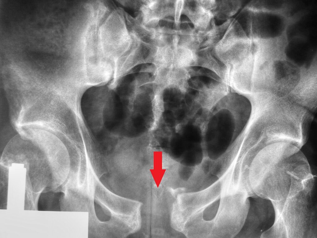
- Standard AP
- Inlet view
- tilt X-Ray beam 40° caudad
- shows posterior displacement
- Outlet view
- 40° cranial beam
- shows superior migration or rotation
CT Scan and Reconstructions
- plan surgical approach
Angiography
- embolisation of bleeding vessels
Treatment
Resuscitation
- fluid replacement
- antishock garment
- embolisation
- direct surgical intervention
- application of Ex Fix can reduce venous and bony bleeding
Provisional Stabilisation
- for fractures that increase pelvic volume
- ie open book (B1) or vertical shear (C3)
- apply ex fix or pelvic clamp percutaneously in emergency room
- External fixation
- 2 pins placed percutaneously in Ilium
- 1 at ASIS,
- 1 at iliac tubercle,
- at ~ 45° to each other
- complete frame as anterior rectangle
- 2 pins placed percutaneously in Ilium
By Type
| Type | Treatment |
|---|---|
| A | symptomatic, mobilisation |
| B1 | Stage 1 . no stabilisation Stage 2+3 . stabilise with External fixateur or anterior plate |
| B2 | most need no stabilisation |
| B3 | displaced bucket handle if LLD less than 1.5 cm . accept . if LLD more than 1.5 cm or pelvic deformity excessive . reduction by ER of hemipelvis with pins in the iliac crest . maintained with anterior frame |
| C | Anterior frame and skeletal traction (supracondylar femoral pin) . ORIF |
Complications
- Non-union / malunion
- Infection
- increased incidence associated with open bowel injury
- 6% incidence
- increased with ilio-inguinal approach
- avoid operations in febrile patients
- use prophylactic antibiotics
- Nerve palsy
- usually peroneal component of sciatic nerve
- 11.2% (17.4% of posterior fractures)
- usually peroneal component of sciatic nerve
- Ectopic bone formation
- ~ 20%
- Thrombo-embolic problems
- urethral injury
- About 1/3 of unstable fractures (13% overall)
- Impotence
- ~ 40%
- Post traumatic osteoarthritis
- 4 – 15% dependent on quality of reduction
Prognosis
- Mortality
- Overall 5 – 20%
- Open fractures up to 42%
- Increasing age
- increased mortality
- Age more than 70 years
- 50% mortality
- Pedestrians
- 50% mortality
- Pregnancy
- 33% foetal loss
- 20 – 40% of females subsequently need caesarean section
Reviewed by Professor Phong Tran, Head of Orthopaedic Surgery, Western Health. Last updated 10 October 2026.