
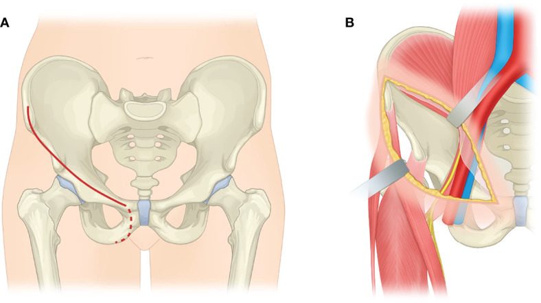
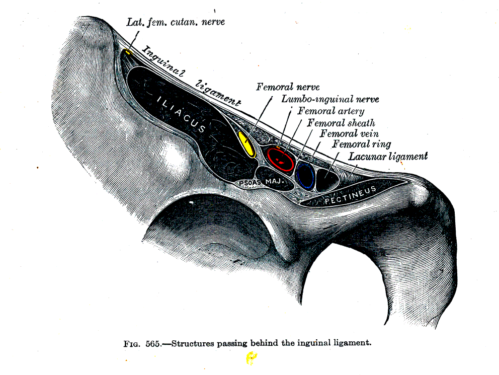
Indications
- exposure of inner surface of pelvis from the sacroiliac joint to pubic symphysis
- anterior & medial surfaces of acetabulum
Contraindications
- does not expose articular surface of acetabulum
Principles
- not for occasional surgeon
- in conjunction with general surgeon
- cadaveric dissection
Position
- supine with greater trochanter at edge of table
- IDC
Landmarks
- ASIS
- Pubic tubercles
Incision
- Curved anterior incision
- Start: 5cm above the ASIS extending medially passing 1 cm above the pubic tubercle
- End: midline
Superficial Dissection
- Dissect through subcutaneous fat in line of the skin incision to expose the aponeurosis of external oblique muscle
- Lateral Cutaneous Nerve of Thigh appears at the lateral edge of the wound & will need to be divided
- Divide
- Aponeurosis of external oblique muscle from superficial inguinal ring to ASIS
- This exposes spermatic cord / round ligament
- Isolate these structures in a sling
- Anterior part of rectus sheath
- Exposes rectus abdominis
- Aponeurosis of external oblique muscle from superficial inguinal ring to ASIS
Deep Dissection
- Divide rectus abdominal muscle transversely 1 cm proximal to its insertion
- Develop a plane using blunt dissection (finger) between pubic symphysis & bladder
- Divide internal oblique & transversus abdominis muscles that form the posterior wall of inguinal canal
- Ligate the inferior epigastric artery at the medial edge of deep inguinal ring
- Exposes the peritoneum & extraperitoneal fat
- Elevate the peritoneum upwards to reveal
- Femoral vessels
- Femoral nerve
- Tendon of iliopsoas
- Isolate these structures & protect with sling, retract medially
- Dissect iliacus off iliac wing
- Exposes the medial surface of acetabulum & superior pubic ramus
Dangers
- Nerve
- Femoral
- Lateral cutaneous nerve of thigh
- Vessels
- Femoral
- Inferior epigastric artery & vein
- Spermatic cord
- Bladder
Reviewed by Professor Phong Tran, Head of Orthopaedic Surgery, Western Health. Last updated 10 October 2026.