
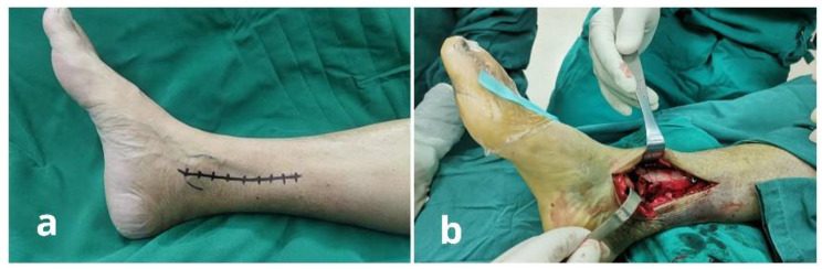
Aims
- Approach the ankle joint via medial incision
Indications
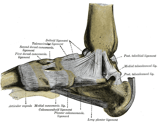
- arthrodesis
- excision of osteochondral fragments from medial side of talus
- r/o loose bodies
Position
- supine
- partial exsanguination
Landmarks
- medial malleolus
Incision
- 10 cm longitudinal incision
- Centred over tip of medial malleolus
- Start: medial surface of tibia
- End: medial cuneiform
Internervous Plane
- None
Superficial Dissection
- Mobilise skin flaps
- Beware
- Long saphenous vein
- Saphenous nerve
- Beware
Deep Dissection
- Divide flexor retinaculum
- Retract Tib Post posteriorly
- Medial Malleolar osteotomy
- Score M.M. longitudinally to ensure correct alignment during closure
- Drill & tap medial malleolus so that it can be reattached
- Incise anteromedial ankle joint to help identify anterolateral junction of distal tibia
- Oscillating saw
- Cut through MM obliquely from top to bottom
- Retract MM with deltoid ligament inferiorly
- Evert foot to expose talar dome
Dangers
- Nerves
- Saphenous
- Veins
- Long saphenous vein
- Tendon
- Tibialis posterior
Reviewed by Professor Phong Tran, Head of Orthopaedic Surgery, Western Health. Last updated 10 October 2026.