
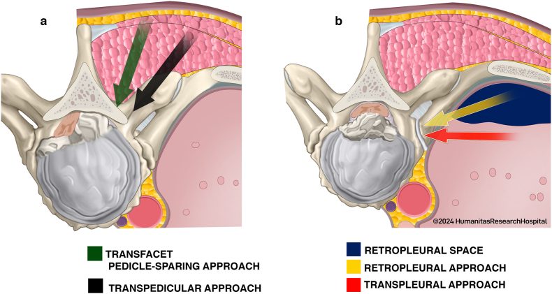
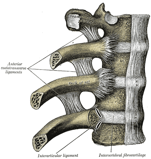
- Unrivalled exposure to anterior portions of vertebral bodies T2 – T12
- Operate in conjunction with Thoracic surgeon
Indications
- Infections TB
- Fusion
- Tumour
- Bone grafting
- Correction of scoliosis
- Dwyer instrumentation & rods
- Correction of kyphosis
Position
- Lateral
- Stabilised with lateral supports
- Patient’s arm placed above the head
- Axillary roll & feel for radial pulse in dependent arm
- Surgeon is behind the patient
- Approach from RIGHT side to avoid aortic arch & aorta
- NG tube to help identify oesophagus
Landmarks
- Tip of scapula
- Spines of thoracic vertebrae
- Inframammary crease
Incision
- L shaped incision over 7th or 8th rib
- Start: 2 fingerbreadths below tip of scapula
- curved forward towards inframammary crease
- curved backward & upward midline between spine & medial border of scapula
Superficial Dissection
- Divide latissimus dorsi in line with skin incision
- Divide Serratus Anterior along the same line, down to the RIBS
- scapula can now be elevated to expose the underlying ribs
- Many vessels are encountered & are cauterised
- Intercostal space or Rib resection
- intercostal space / rib to be resected depends on the location of pathology
- 5th intercostal space
- T2 – T10
- 6th intercostal space
- T10 – 12
- Cut down to rib with cutting diathermy
- Cut the periosteum on the upper border of the rib
- If resecting the rib
- Resect the posterior ¾ of the rib as far posterior as necessary
- Insert rib spreader to hold ribs apart
Deep Dissection
- Ask Anaesthetist to DEFLATE the lung
- Gently retract it anteriorly with moist lap pads to protect it
- Identify the Oesophagus
- Incise the pleura over the oesophagus so that it can be mobilised
- Ligate / tie off intercostal vessels that are necessary
- Retract & hold oesophagus medially with 2 penrose drains
- vertebral bodies are now exposed
- If need to reach T11 –T12
- Part of diaphragm may need to be resected
- Remove arcuate ligament from its origin on transverse process of L1
Dangers
- Intercostal vessels
- Lungs
- Inflate lungs every 30 minutes to help prevent microatelectasis postop
Reviewed by Professor Phong Tran, Head of Orthopaedic Surgery, Western Health. Last updated 10 October 2026.