
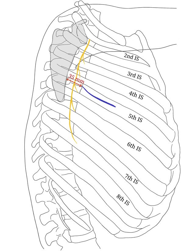
Decompression of neural elements when anterior neural compression has been documented.
Indications
- traumatic
- decompression for documented neurocompression 2º to bone or disc fragments anterior to dura
- infectious
- open biopsy for diagnosis, debridement & anterior strut grafting
- degenerative
- removal of disc herniation with interbody fusions
- neoplastic
- extradural metastatic disease
- 1º vertebral body tumour
- deformity
- kyphosis/scoliosis
- (congen/acquired/idiopathic)
Use
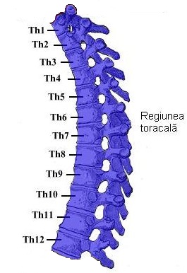
- provides direct access to vertebral bodies T2 to T12
- Midthoracic vertebral bodies are best exposed by this approach
- Campbells
- L-sided thoracotomy preferred, heart may be retracted anteriorly
- R-sided thoracotomy used by some for approaching upper Tx spine to avoid subclavian & carotid arteries in L superior mediastinum, liver may present significant obstacle to exposure inferiorly
- Hoppenfeld
- right side easier
Prerequisites
- Expertise at approach
- Thoracic surgeon able to deal with hazards in area
Positioning
- lateral decubitus position with right side down
- inflatable beanbag with table flexed to ↑ exposure
- Arm on operating side lateral to head
- Ax roll
- Attend to pressure points
- NG tube
Incision
- Make incision 2 levels above
- Eg. For L5 cut at L3
- divide latissimus dorsi & serratus anterior
- Expose rib subperiosteally
- (if multiple levels are involved, rib at upper level of proposed dissection should be removed) – inferior angle scapula is landmark
Procedure
- Use electrocautery to maintain haemostasis during exposure
- Resect posterior 3/4 of rib
- Take care to identify & preserve intercostal nerve lying along inferior aspect of rib as it localizes neural foramen leading into spinal canal
- Insert rib spreader
- Ask gas man to deflate lung
- Divide pleura exposing lung
- Retract lung anteriorly using lap pads
- Identify oesophagus if on R side by palpating NG tube
- Incise pleura over lateral side of oesophagus so can retract oesophagus & reflect pleura exposing anterior spine, usually one vertebra above & one below involved segment, to allow adequate exposure for debridement & grafting
- Identify & ligate segmental vessels & azygous vein crossing surgical field
- Exposure from L is more difficult as need to retract aorta & ligate both R & L segmental arteries for full exposure
- Carefully reflect periosteum overlying spine with elevators to expose involved vertebrae
- Use small elevator to clearly delineate pedicle of vertebrae & Kerrison rongeur to remove pedicle, thus exposing dural sac. Identify disc spaces above & below vertebrae & incise annulus. Remove disc material using rongeurs & curettes. entire cross section of vertebral body is thus developed, & anterior margin of neural canal is identified with posterior longitudinal ligament lying in slight concavity on back of vertebral body. Expose sufficient segmental vessels & disc spaces to accomplish intended procedure—usually corpectomy & strut grafting
Complications
- Atelectasis, Lung injury, pneumothorax, haemothorax
- Vessel injury, Intercostals vessel damage
- Visceral damage: Oesophagus
- Nerve injury
- Cord injury
- Clicking of scapula over operative site
Reviewed by Professor Phong Tran, Head of Orthopaedic Surgery, Western Health. Last updated 10 October 2026.