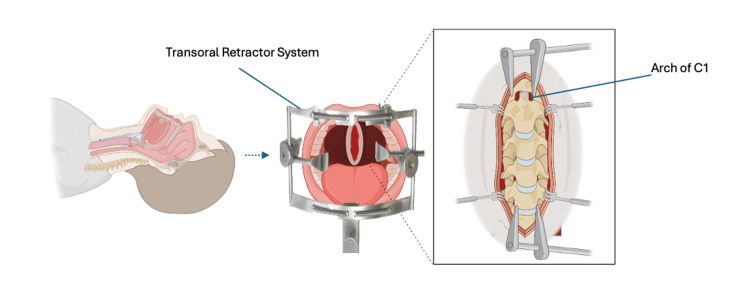
 Transoral approach to C1 and C2, with a self retaining retractor holding the mouth open and a midline incision through the posterior pharyngeal wall exposing the anterior arch of C1 and the upper odontoid. Image from Sadeh M, Mansour H, Iqbal J, Gonzales-Portillo G, Souter J, Neckrysh S, Callahan N, Atwal G. Transoral Approach to the Craniovertebral Junction, A Case Series of Three Rare Pathologies. Cureus. 2025, 17(10), e94046. CC BY 4.0.
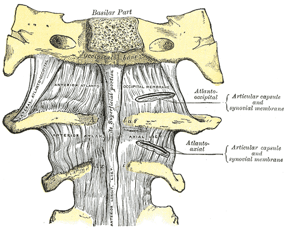 Anterior atlanto-occipital membrane and atlantoaxial ligaments, the structures exposed behind the posterior pharyngeal wall in the transoral approach. Image by Henry Vandyke Carter, Gray’s Anatomy (1918), Wikimedia Commons, public domain.
- fiberoptic nasotracheal intubation & NG tube
- Mayfield head rest with head slightly extended
- cleanse oral cavity with chlorhexidine
- preop ABx = IV Ancef & Flagyl
Landmarks
- anterior tubercle on atlas (ALL & longus colli muscles attached)
Superficial Dissection
- inject posterior oropharynx with 1:200,000 epinephrine
- midline 3 cm vertical incision centred on anterior tubercle
- expose anterior arch subperiosteally
Reviewed by Professor Phong Tran, Head of Orthopaedic Surgery, Western Health. Last updated 10 October 2026.
