
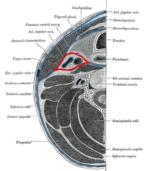
Position
- Place appropriately prepared patient supine on operating table with a Mayfield headrest
- Administer IV antibiotics
- Turn head away from side of approach, usually towards right
- Elevate table 30°
- decreases venous bleeding
- Prepare skin & drape patient, exposing neck from jaw to jugular notch
Landmarks
- Hard palate – arch of atlas
- C2-3 – lower border of mandible
- C3 – hyoid
- C4-5 – thyroid cartilage
- C6 – cricoid cartilage & carotid tubercle
- sternocleidomastoid
Incision
- Transverse incision at level of fusion
- From posterior border of SCM to midline
- It is possible to access up to three levels via one transverse incision, but if wider access is required, less cosmetically pleasing incision can be made along anterior border of sternocleidomastoid
Internervous Plane
- Platysma split
- Facial nerve proximally
- SCM
- Accessory spinal nerve
- Strap muscles
- Segmental innervation C1-3
Superficial Dissection
- Divide fascia over platysma in line with skin incision
- Split fibres of platysma in their line using blunt dissection
- Locate SCM (spinal accessory nerve)
- & divide fascia along its anterior border, retract SCM laterally
- Retract strap muscles medially (segmental innervation)
- Sternohyoid
- Sternothyroid
- Locate carotid sheath, & divide pretracheal fascia along medial aspect of sheath
- Common carotid artery
- Vein
- Vagus nerve
- Retract carotid laterally
- Retract midline structures medially
- trachea & oesophagus & thyroid gland
- Dangers
- superior & inferior thyroid arteries
- connect the carotid sheath to midline structures
- middle thyroid vein
- These may be divided if required
- superior & inferior thyroid arteries
Deep Dissection
- Cervical vertebrae are visible covered by
- Anterior longitudinal ligament
- Longus colli muscle
- sympathetic chain
- Prevertebral fascia
- Divide prevertebral fascia vertically, in midline down to bone
- Reflect subperiosteally
- prevertebral muscles & sympathetic chain & ALL
- to expose the anterior vertebral body
Dangers
- Nerves
- Recurrent laryngeal nerve
- Right
- may cross transversely
- Left
- Nerve is more constant in its course
- May be damaged during deepest layer of approach
- Right
- Sympathetic nerves & stellate ganglion
- Horner’s syndrome
- Recurrent laryngeal nerve
- Vessels
- Carotid sheath & contents
- Vertebral artery
- Superior & inferior thyroid arteries
Reviewed by Professor Phong Tran, Head of Orthopaedic Surgery, Western Health. Last updated 10 October 2026.