
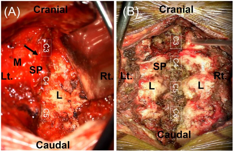
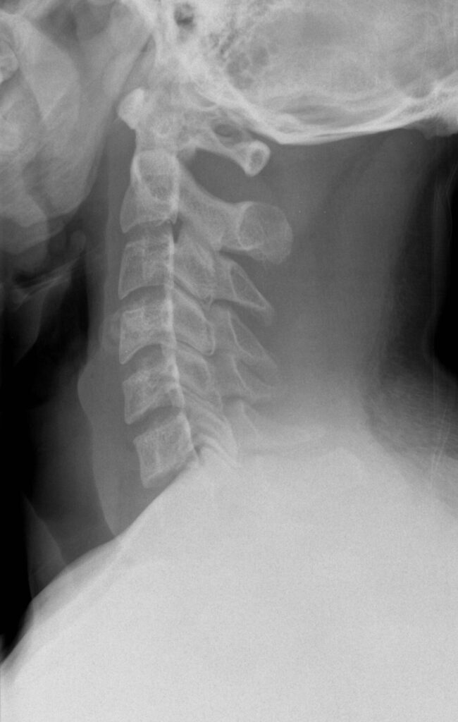
Indications
- Entire cervical spine
- Posterior spinal fusion
- Herniated discs
- Tumours
- Facet joint dislocations
- Nerve root exploration
Position
- prone
- mayfield head rest
- flex head a few°
- Prep & Drape
Landmarks
- Spinous processes
- C2
- C7
- T1
- Identify level with Spinal needle & II
Incision
- Straight longitudinal midline incision
Internervous Plane
- Left & right paracervical muscles
- Supplied segmentally by posterior rami
Superficial Dissection
- Continue incision down to spinous processes
- Reflect paraspinal muscles subperiosteally using a Cobb Elevator
- Carry the dissection laterally to reveal the Lamina, Facet Joints, & Transverse Processes
Deep Dissection
- Identify Ligamentum Flavum & remove it from the Superior edge of the Inferior Lamina
- Perform a laminotomy / laminectomy
- Expose blue white dura & epidural fat
- Gently retract the spinal cord medially to expose the vertebral body, disc space
- Watching for the thin epidural veins
Dangers
- Nerves
- Spinal cord
- Nerve roots
- Vessels
- Venous plexus in the cervical canal
Reviewed by Professor Phong Tran, Head of Orthopaedic Surgery, Western Health. Last updated 10 October 2026.