
Indications
- Distal humerus ORIF
- Exploration of radial nerve
Position
- Supine
- Arm board
Landmarks
- Biceps
- Anterior flexion crease of elbow
Incision
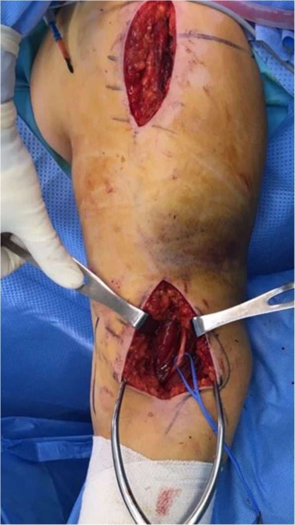
- Curved longitudinal incision along lateral border of biceps
- Start: 10 cm above elbow crease
- End: just above elbow crease
Internervous Plane
- Brachialis
- Brachioradialis
- Radial nerve
Superficial Dissection
- Beware lateral cutaneous nerve of forearm
- Identify lateral border of biceps & retract medially
- Develop the plane between brachialis & brachioradialis
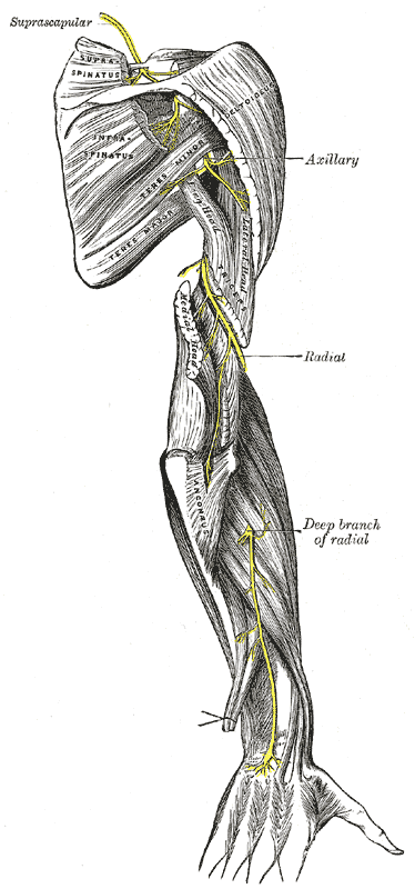
- Identify the radial nerve near the elbow & follow it up to where it pierces the lateral intermuscular septum
- Isolate & retract radial nerve laterally with vascular loops
Deep Dissection
- Incise along the lateral border of brachialis down to periosteum & bone
Dangers
- Nerves
- Lateral cutaneous nerve of forearm
- Radial nerve
Reviewed by Professor Phong Tran, Head of Orthopaedic Surgery, Western Health. Last updated 10 October 2026.