
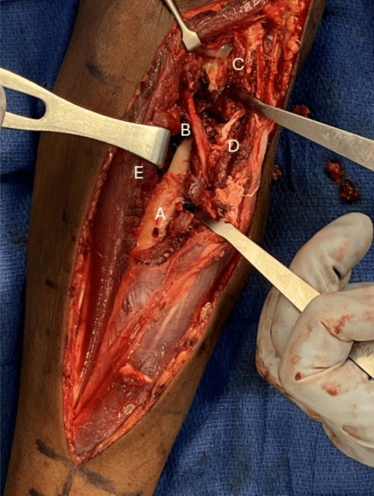
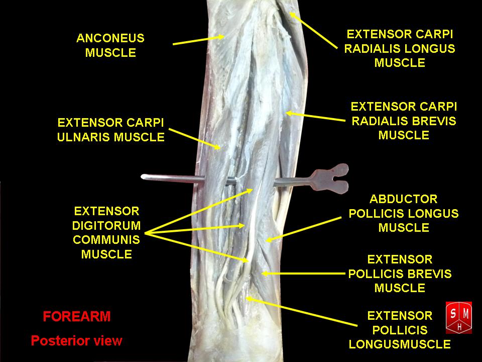
Thompson Approach
Position
- Supine
- Forearm pronated
Landmarks
- Lateral epicondyle of humerus
- Lister’s tubercle
Incision
- Straight longitudinal incision
- Start anterior to lateral epicondyle
- End just ulnar to Lister’s tubercle
- Use whatever part that is necessary
Internervous Plane
- Proximally
- ECRB radial nerve
- EDC PIN
- Distally
- ECRB radial nerve
- EPL PIN
Superficial Dissection
- Deep fascia
- Identify interval between ECRB & EDC distally
Deep Dissection
- Proximal 1/3
- Radius is covered by supinator
- PIN is between superficial & deep heads & arises 1cm proximal to distal edge
- Identify & preserve PIN
- Mobilise the nerve by carefully dissecting it through substance of supinator
- Supinate forearm
- Detach Supinator from origin subperiosteally
- Middle 1/3
- Radius covered by
- APL
- EPB
- Radius covered by
- Distal 1/3
- Identify interval between EPL and ECRB
- Radius is covered by supinator
Dangers
- PIN
Reviewed by Professor Phong Tran, Head of Orthopaedic Surgery, Western Health. Last updated 10 October 2026.