
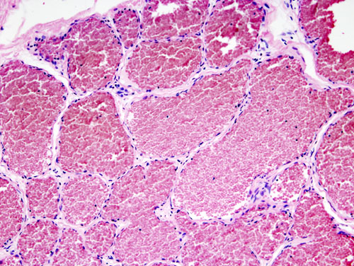
Definition
- haemangiomas are benign lesions characterised by vascular spaces lined with endothelial cells, 50% in spine, 20% in skull
Presentation
- 40-50 peak incidence
- usually asymptomatic (incidental finding)
- neurological symptoms or back pain
Investigations
- X-ray
- vertebra
- thickened vertical trabeculae secondary to erosion of horizontal trabeculae
- skull
- radiating wheel spoke
- vertebra
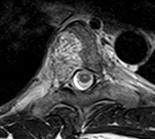
- CT
- polka dot appearance
- MRI
- T1 low, T2 high
- Bone scan
- repeat local staging at hot spots
- Biopsy
Treatment
Non-operative
- If asymptomatic – observe
- Radiation
- spine
- Embolisation
- either as Treatment or preop
Operative
- Excision & bone graft
- always preop embolisation
Reviewed by Professor Phong Tran, Head of Orthopaedic Surgery, Western Health. Last updated 10 October 2026.