
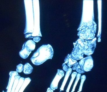
Also Known As
- dysplasia epiphysealis hemimelica
- Trevor’s disease
Epidemiology
- Very rare
Aetiology
- Non familial developmental disorder
- Benign
Presentation
- Manifests in young children
- Unilateral enlargement of an epiphysis
- Most commonly affects lower femur, upper tibia & talus
- If affecting the proximal femoral epiphysis can be confused with Perthes disease
- Can cause varus or valgus deformity
Histology
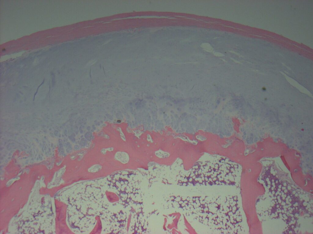
- resembles an osteochondroma, with a cartilage cap of disorganised cartilage
Treatment
- surgical excision
Reviewed by Professor Phong Tran, Head of Orthopaedic Surgery, Western Health. Last updated 10 October 2026.