
- A benign tumour of cartilaginous origin with a predilection for the epiphysis in skeletally immature patients
- “Codman’s tumour”
- Epiphyseal chondromatous giant cell tumour
Definition
- Benign cartilage lesion
- But aggressive/ frankly malignant behaviour reported
- Arises in secondary ossification centre
- Develops until physis closes
Epidemiology
- Rare cartilage tumour
- < 1% of all bone tumours
- Found in 2nd decade with peak at 20 years
- M:F – 1.5:1
- Appears in secondary ossification centre
- Epiphysis of long bones (cf. Enchondromas in meta-diaphysis)
- Proximal humerus (Codman’s tumour)
- Around knee
- Distal femur
- Proximal tibia
- Epiphysis of long bones (cf. Enchondromas in meta-diaphysis)
- On rare occasions seen in
- Older patients
- Odd locations such as spine & flat bones
Clinical Features
- Adolescent or young adult
- Pain & swelling
- Can restrict ROM of joint
- Confused with intra-articular pathology
- Can alter the epiphyseal development in young children
Investigations
- X-ray
- Well-demarcated lucent defect (ie. radiolucent with sclerotic margin)
- Epiphyseal in location & eccentric
- May extend into metaphysis
- 25-50% have punctate calcification
- 25% have popcorn or chicken-wire calcification
- < 10% have periosteal reaction
- Cortical bone intact or expanded
- Bone Scan
- Very hot
- MRI
- Oedema seen in surrounding tissues
Pathology
- Gross
- Gritty/ Greyish pink material
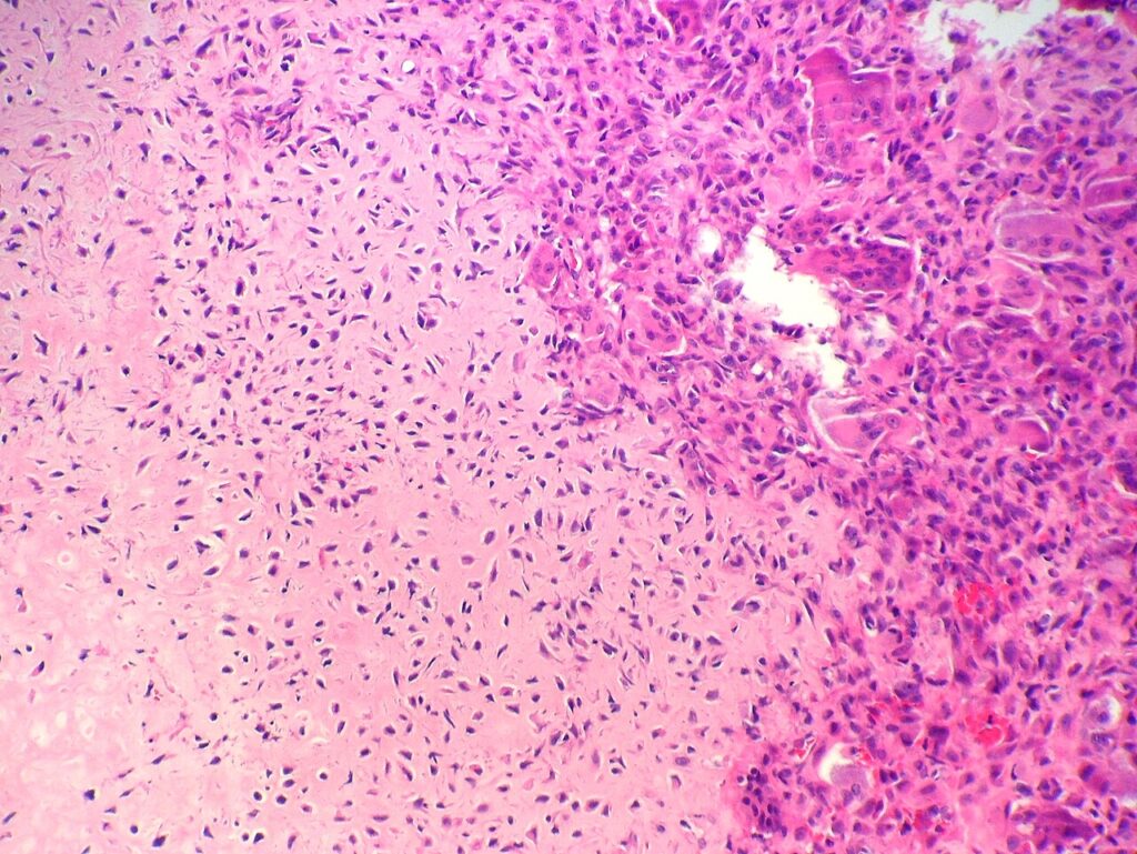
- Microscopic
- Highly cellular & undifferentiated tissue
- Sheets of Chondroblasts
- Round & Polygonal cells (plump or like fried eggs)
- Bluish cytoplasm
- Occasional multinucleated cells of osteoclast type
- Small areas of cartilaginous matrix & extracellular calcification present
- Chicken wire calcification
- Atypical cartilage matrix
- Stains positive for S100
- Chondroid Matrix
- Differentiate from
- GCT by absence of typical spindle stromal cells of GCT
- 20% are cystic & haemorrhagic similar histologically to ABC
Differential Diagnosis
- GCT
- OCD
- Brodies abscess
- Clear Cell Chondrosarcoma
- Enchondroma
- Fibrous dysplasia
Treatment
- Difficult to eradicate due to anatomic position without damaging joint or physis
- Intralesional curettage & bone grafting
- usual approach
- Often access through growth plate as patient at or near end of growth
- Ie. preserve the joint rather than growth plate
- If recurrence occurs then wide resection procedure of choice
Intra-lesional Curettage
- Technically more feasible
- Intra-articular exposure required
- Local Adjuvant Therapy
- Phenol
- Liquid Nitrogen
- Avoid damage to physis if possible
- Bone graft
- Recurrence may occur
- Usually cured with 2nd curettage
Wide Resection
- Lower recurrence rate
- Usually not possible without loss of function
- Indicated for 2nd recurrence
- There is incidence of pulmonary lesions which are rimmed with bone & should be excised
Prognosis
- The recurrence rate with chondroblastoma alone is 20% at 3 years
- If the tumour has an aneurysmal component, the risk of recurrence is higher
- Follow-up care is necessary because recurrence is common
- Radiographs should be repeated every 6-12 months after excision for ~2 years
Reviewed by Professor Phong Tran, Head of Orthopaedic Surgery, Western Health. Last updated 10 October 2026.