
Definition
- Small (< 1cm diameter of central nidus), solitary, benign, painful bone-forming tumours
Epidemiology
- Relatively common
- 10% of all primary bone tumours
- Classically children & adolescents
- Aged 5-25 years
- M:F – 2:1
- Most common site is lower extremity long bones (60-70%) usually near diaphysis
- Femur 30%
- Femoral neck & Intertrochanteric area most common femoral location
- Tibia 25%
- Foot 10%
- Talus & Calcaneum most common in foot
- Humerus 10%
- Forearm 3%
- Hand 10%
- Scaphoid most common in wrist
- Spine
- Can occur in spine (Osteoblastoma more commonly) in posterior elements
- Femur 30%
- Not infrequently located at the site of previous trauma
- ? pathogenesis associated with trauma
Clinical Features
- Characteristic pain
- Intense, unrelenting
- Chronic pain months/ years
- Nocturnal
- Due to prostaglandin production
- Relieved by aspirin (narcotics often unhelpful)
- Local swelling
- Exquisitely tender
- Mild Leucocytosis may be present
- If intra-articular/ juxta-articular may present as synovitis of joint
- Spinal lesions will show irritative scoliosis often
Radiology
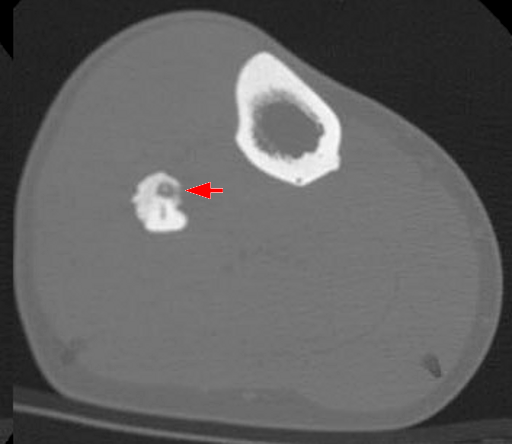
X-ray
- Cortical location in long bone
- Central lucent zone (Nidus)
- Increased surrounding bone density with sclerosis
- Fusiform shaped
- May obscure nidus
- Marked periosteal reaction may be present
- Subperiosteal location = marked sclerosis
- Endosteal location moderate sclerosis
- Subarticular location = often no sclerosis
- Nidus can be obscured by the surrounding sclerosis
- Medullary
- Four features (~ 50% show all 4)
- Sharply round or ovoid
- < 1cm
- Homogenous dense centre
- 1mm peripheral radiolucent zone
- Four features (~ 50% show all 4)
- Differential on XR includes
- Stress fracture
- Osteomyelitis
CT Scan
- Demonstrates nidus better
- Location in cortex clearer
- Thin sections 1-2mm
- Low attenuation nidus
- Central mineralisation
- Surrounding endosteal & periosteal sclerosis
Bone Scan
- Help with diagnosis & localisation of the tumour
- Extensive ↑ uptake
- Avidly take up the isotope due to osteoblastic activity in nidus
- Also develops intense surrounding reaction
- Can help localise intraoperatively
Pathology
Gross
- Well demarcated cherry red nidus with gritty consistency
- Surrounding dense bone with periosteal reaction & thickening
Microscopic
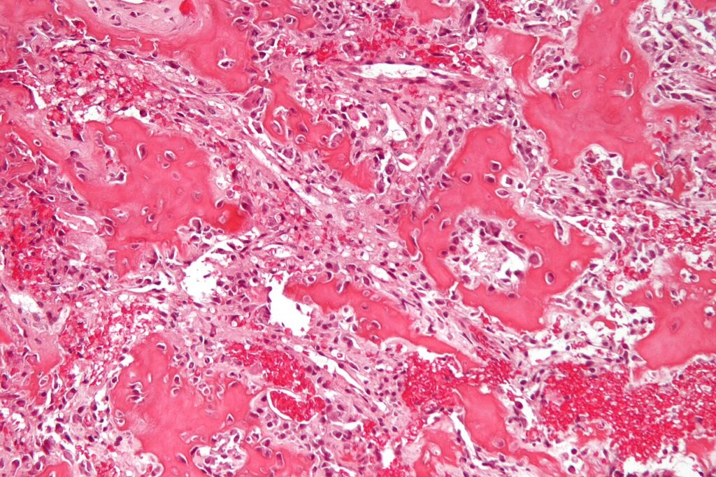
- Maze of small spicules of immature bone
- Haphazard
- Delicate trabeculae of osteoid
- Rimmed by numerous osteoblasts
- Enclosed in vascular spindle cell stroma
- Numerous vascular channels & capillary network
- Rich nerve fibres
- Giant cells may be present
- More mature lesions stroma sparsely cellular with intervening vascular spaces
- No chondroid elements
Differential Diagnosis
- Osteoblastoma
- Brodie’s abscess
- Stress fracture
- Bone Island
- Eosinophilic Granuloma
- Osteosarcoma
- Osteoma
Treatment
- Generally remove the nidus
- (May heal spontaneously but takes long time)
- En Bloc Excision
- Gold standard
- Ensures all of lesion excised
- Weakens bone
- Need graft & protection
- Send for intraoperative FFS & XR
- “Burr Down” Technique
- Excellent results
- Intraoperative CT guidance
- Direct incision over lesion
- Shave cortex off with high speed burr to reactive bone
- Scoop nidus out once hit hypervascular zone & sent for FFS
- Burr 2mm zone out
- Can leave strong reactive bone behind
- Percutaneous Guided Reaming
- CT-guided in XR suite
- Doesn’t produce tissue for histology
- Radiofrequency Ablation
- CT-guided
- No tissue for histology
- Death occurs by thermal ablation
- Uncertain results
- Methods of localisation include
- Image Intensifier
- CT-guidance
- Radioisotope probe
- Intraop tetracycline fluorescence
- If complete excision performed then recurrence very rare
- However incomplete excision not uncommon & often leads to recurrence & failure of pain relief
OsteoBlastoma
Reviewed by Professor Phong Tran, Head of Orthopaedic Surgery, Western Health. Last updated 10 October 2026.