
Epidemiology
- Atlas fracture is rare in children
- Axial load from a fall onto the vertex or a diving injury
- Burst (Jefferson) pattern, often through cartilaginous synchondroses in young children
Anatomy
- Three primary ossification centres, the anterior arch and two neural arches
- Anterior arch may be unossified at birth, ossifying in the first year
- Posterior synchondrosis fuses around 3 to 4 years
- Anterior (neurocentral) synchondroses fuse around 7 years
- Unfused synchondroses appear as lucent lines with smooth corticated margins
- Congenitally incomplete or bifid posterior arch mimics fracture
- Pseudospread, lateral masses overhanging C2 on the open mouth view, as C1 outgrows C2 in early childhood
Clinical Features
- Neck pain and reluctance to move the neck
- Torticollis
- Neurological deficit uncommon, as the burst pattern widens the canal
- Look for associated head injury and other cervical injuries
Investigations
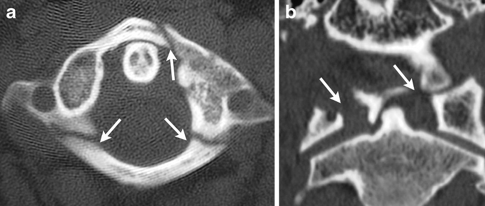
- Plain radiographs are difficult to interpret in young children
- CT defines the fracture and distinguishes it from synchondrosis
- MRI assesses the transverse atlantal ligament and soft tissues
- Lateral mass overhang is a less reliable marker of transverse ligament rupture than in adults
Management
- Most treated non-operatively and heal well
- Hard collar for stable injuries
- Halo or Minerva immobilisation for unstable or displaced injuries
- Young children need more halo pins at lower insertion torque because the skull is thin
- CT before halo application guides pin sites away from sutures and thin bone
- C1 to C2 fusion is rare, for transverse ligament instability or failed non-operative care
Complications
- Halo pin site infection and loosening
- Dural puncture from pins
- Persistent instability from a missed ligamentous injury
Reviewed by Professor Phong Tran, Head of Orthopaedic Surgery, Western Health. Last updated 10 October 2026.