
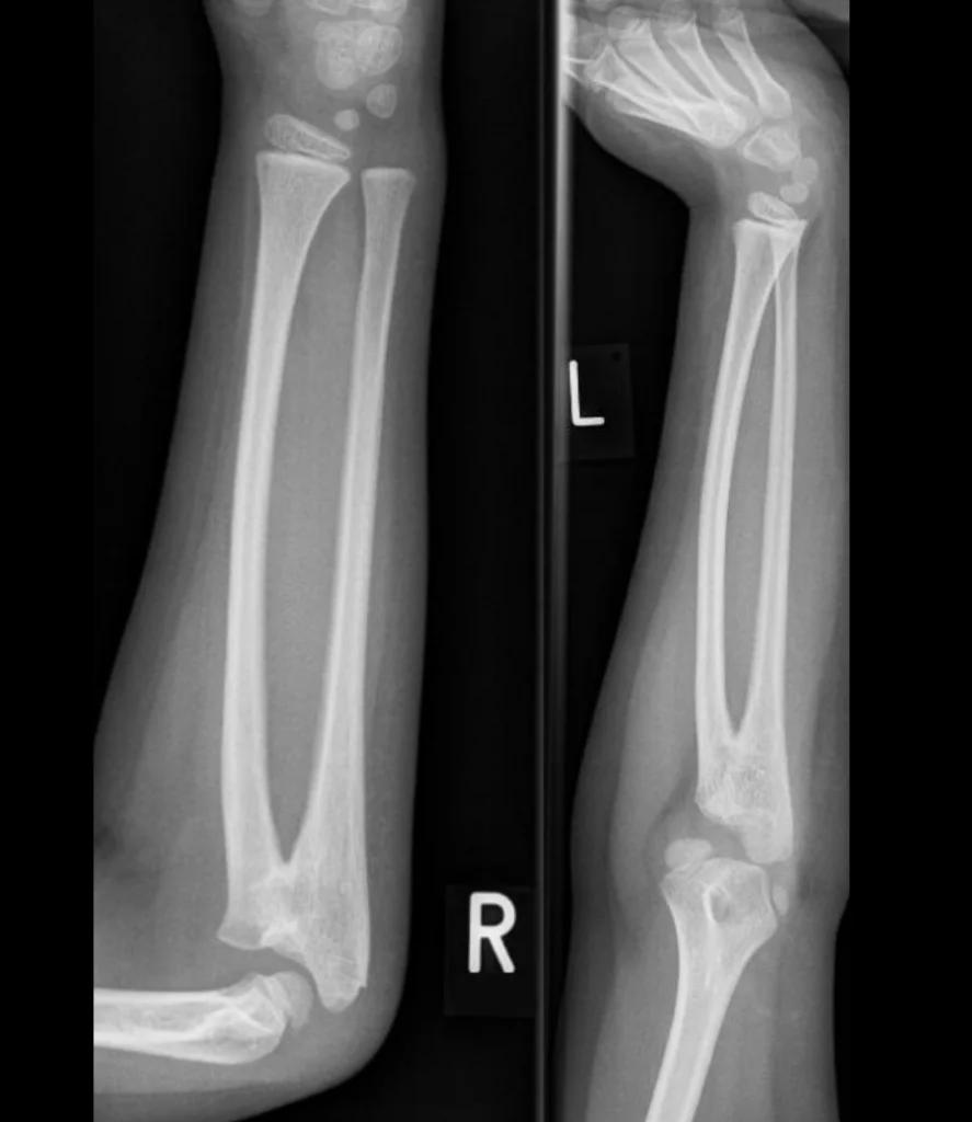
Definition
Abnormal bony connection between the radius and ulna from birth.
Anatomy
—
Epidemiology
Approximately 60% bilateral pathology
In some cases appears to be associated with autosomal dominant pattern
Even distribution between males and females (some sources state slightly more common in males 3:2)
30% of cases involved with syndromes – Klinefelter syndrome, XXXY syndrome, Apert syndrome, Crouzon syndrome, carpenter syndrome, arthrogryposis, Holt-Oram syndrome, Williams syndrome.
Aetiology
Abnormality of longitudinal segmentation
Radius and ulna initially one anlage which then divides from distal to proximal
Failure of this separation as distal to proximal segmentation occurs causing synostosis
Pathology
—-
Natural History
Congenital pathology. Does not progress.
History
Frequently unnoticed in first years of life
Pain free
Can present with issues grasping or holding items due to reduced rotational movement – carrying items, keyboard, catching balls and feeding self
Parents or caregivers (teachers, sports coaches) often first to report issue
Examination
Commonly no obvious deformity, however can have observable bony deformity if severe.
Varying amounts of flexion/extension range of motion depending on extent of synostosis
Pronation/supination range of movement most notably restricted
Forearm shortening and decreased carrying angle can be appreciated
Most common position of fixed pronation is 30 degrees
Compensatory mechanism: shoulder abduction/adduction
Wrist hyper mobility
Differential Diagnosis
—
Classification
Cleary and Omer classification
| Type | Description |
|---|---|
| 1 | Fibrous synostosis with a normal, reduced radial head |
| 2 | Osseous synostosis with a normal, reduced radial head |
| 3 | Osseous synostosis with a hypoplastic, posteriorly dislocated radial head |
| 4 | Short osseous synostosis with an anteriorly dislocated, mushroom shaped radial head |
Original publication Cleary JE, Omer GE Jr. Congenital proximal radio-ulnar synostosis. Natural history and functional assessment. J Bone Joint Surg Am. 1985;67(4):539-45.
Investigation
X-ray. Further imaging rarely indicated if management is non-operative. CT can assist with extent of synostosis and 3D modelling for operative management.
Treatment
Majority of patients managed conservatively – if functional deficit does not significantly impact ability to undertake activities of daily living. This is more common in unilateral disease.
Operative:
Synostosis excision with soft tissue interposition
Forearm de-rotational osteotomy through synostosis or distal
Forearm de-rotational osteotomy with frame
References
Reviewed by Professor Phong Tran, Head of Orthopaedic Surgery, Western Health. Last updated 10 October 2026.