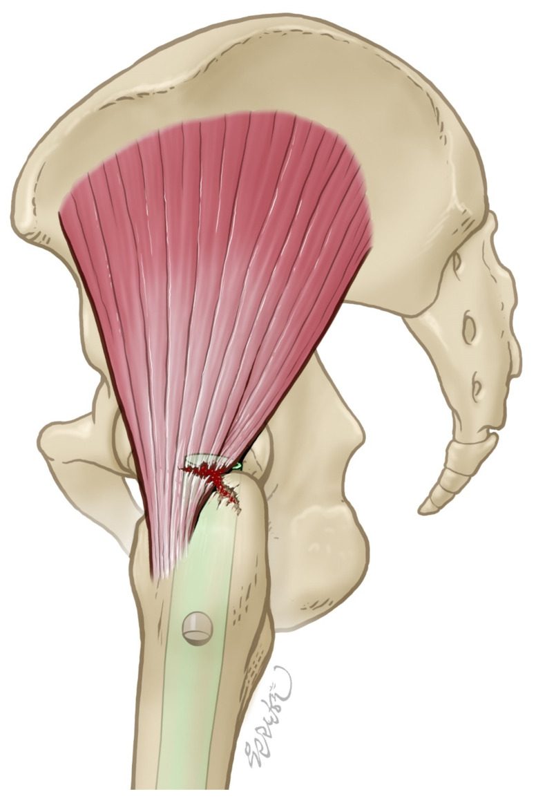
 Drawing of an antegrade nail entering at the tip of the greater trochanter through the gluteus medius insertion, where the abductor tendon can be torn at the entry point. Image from Yoon Y, Lee H, Lam KHS, Lee M, Lee J, Hwang J. The Sonographic Evaluation of Abductor Injury After Intramedullary Nailing for the Hip Fractures. J Clin Med. 2025, 14(15), 5498. CC BY 4.0.
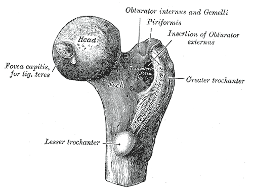 Upper end of the right femur from behind, showing the greater trochanter and trochanteric fossa used as nail entry points. Image by Henry Vandyke Carter, Gray’s Anatomy (1918), Wikimedia Commons, public domain.
Position
- supine
- traction table
- leg adducted
- position checked with II
Landmarks
- greater trochanter
- ASIS
- Shaft of femur
- Patella
- Mark entry point using guide wire in AP & lateral planes
Incision
- Longitudinal incision centred on point intersecting
- Midshaft of femur
- Perpendicular line from ASIS
Internervous Plane
- Split of Gluteus maximus & medius
Superficial Dissection
- Subcutaneous fat & fascia
- Using blunt dissection
- Split the fibres of gluteus maximus & medius
Deep Dissection
- Feel for tip of greater trochanter
Reviewed by Professor Phong Tran, Head of Orthopaedic Surgery, Western Health. Last updated 10 October 2026.
