
- Created by Godfrey Hounsfield in 1972. He won a Nobel prize in 1979
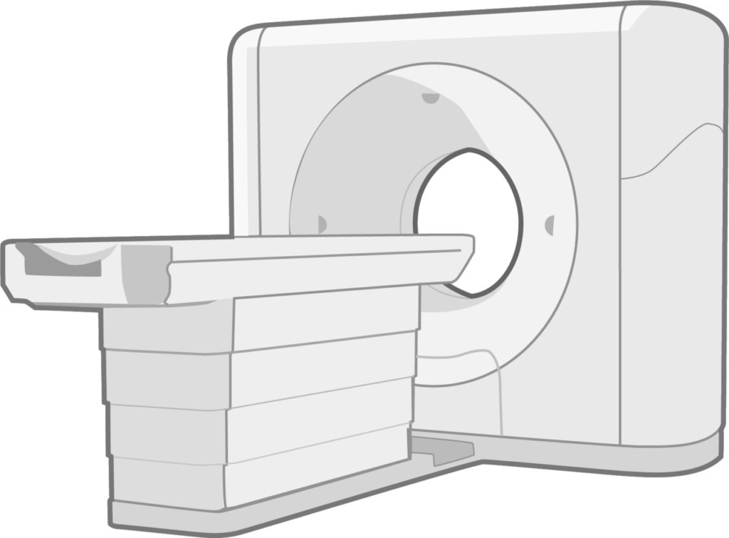
- CT image is created by revolving an XR tube around the patient at high speed, opposite a stationary ring of detectors of very high sensitivity.
- Because of the sensitivity of the detectors a wide range of densities can be obtained
- XR beam is fan shaped
- range of densities in a normal film is around 20. In a CT image it is 2000
- Hounsfield units
- This is a measure of attenuation in CT scans
- Air -1000 units
- Water 0 units
- Cortical bone +1000 units
- range of Hounsfield units displayed can be manipulated by choosing a particular window width. Certain tissues can be highlighted by the appropriate window width; hence “bony windows” & “soft tissue windows”
Plane of imaging
- image is obtained in the axial plane, but images can be reconstructed in any desired plane
Reviewed by Professor Phong Tran, Head of Orthopaedic Surgery, Western Health. Last updated 10 October 2026.