
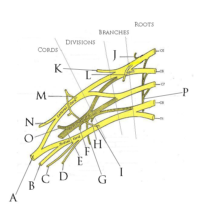
Incidence
- Most common birth palsies
- 1/1000 live births » 1 in 10 of these = permanent impairment
- Increased risks
- high birth weight
- Prolonged labour
- Breech
- Shoulder dystocia
Aetiology
- Lateral flexion of head on trunk
- Forceps may contuse plexus directly
- May occur after CS
Pathology
- Most – at level of neural foramen or groove of transverse process
- Effect varies with force (lower plexus takes less force to disrupt)
- Stages
- Mild
- perineural oedema, haemorrhage
- rapid & complete recovery
- Moderate
- some nerve fibres disrupted, intra & extraneural bleeding
- recovery slow + incomplete
- Severe
- avulsion of trunks or roots
- worst prognosis but some recover
- Incomplete recovery »
- muscle contractures & secondary skeletal changes
- Most commonly medial rotation & adduction of shoulder
- (contractures of subscapularis, pec major, teres major, short head biceps)
- Severe cases
- posterior subluxation or dislocation shoulder
- Flattening of humeral head
- Retroversion of humeral neck
- Glenoid fossa shallow
- Scapula high
- Mild
Classification
| Type | Roots | Features | Frequency |
|---|---|---|---|
| Upper (Erb-Duchenne) | C5, C6 | Weak deltoid, external rotators, biceps, brachialis, brachioradialis and supinator. Waiter’s tip posture with the shoulder adducted and internally rotated, forearm pronated and wrist flexed. Minimal sensory loss, later elbow flexion contracture | Commonest |
| Complete | C5 to T1 | Flaccid paralysis of the whole limb, with or without vasomotor changes giving a marbled hand | Second most common |
| Lower (Klumpke) | C8, T1 | Weak wrist and long finger flexors and intrinsics. Poor hand function with good shoulder and elbow function | Least common |
Diagnosis
- Absence of active movement in the newborn
- Moro reflex absent in affected limb (Grasp reflex lost in complete or lower injuries)
- T1 may be affected » Horner’s (bad prognostic sign)
- Phrenic nerve may be affected » raised hemidiaphragm
Differential
- Pseudoparalysis
- Delivery fractures
- clavicle common (5% associated with obstetric palsy)
- Midshaft humerus
- Dislocation
- shoulder or elbow is rare
- Osteomyelitis / septic shoulder
- Includes E-coli & group B strep
- Delivery fractures
- Arthrogryposis
Management
- Most recover in 1st 3 months
- Use passive ROM 3 – 4 times per day to avoid contractures
- Surgical exploration of plexus
- only after 3 months trial observation
- ± consider electromyography
- Supraclavicular approach
- Nerve reconstruction – graft or repair (sural n)
- Neurotization – (re-routing other nerves)
- Upper plexus = better results than lower
Late Deformity
- Aim to compensate for the fixed adduction & internal rotation
Soft Tissue
- Open reduction shoulder
- Hold temporarily with K-wire
- Shoulder spica
- SEVER release
- Release or lengthen pec major & subscapularis
- Improves range of ER & abduction
- Release or lengthen pec major & subscapularis
- L’Episcopo procedure
- Transfer teres major to more lateral position
- With lateral dorsi » improves ER & abduction power
- lengthen brachialis & biceps
- if elbow fixed flexion is compensating for residual shoulder deformity
- pronator teres lengthening
- Fixed pronation forearm
- osteoclasis of radius may also be needed
- Fixed pronation forearm
Bone
- External rotation humerus
- After 6 years old
- But by then = ~40 % chance of posterior dislocation (therefore reduce earlier)
Reviewed by Professor Phong Tran, Head of Orthopaedic Surgery, Western Health. Last updated 10 October 2026.