
- Craniofacial skeleton
- derived from neural crest & brachial arches
- intramembranous except for base of skull which is endochondral
- Axial skeleton
- derived from sclerotome of somites
- largely endochondral
- Appendicular skeleton
- derived from lateral plate mesoderm that contributes to the formation of limb buds
- largely endochondral
Intramembranous
- Directly from mesenchymal condensations
- Begins at end of embryonic period
- Mesenchymal cells differentiate into osteoblasts & lay down osteoid
- eg. Cranial vault, facial bones, clavicle
Endochondral
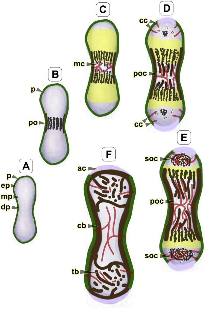
- mesenchymal (blastemal) condensation changes into a cartilaginous miniature precursor at end of embryonic period
- mesenchymal elements begin to chondrify at ~ 6w of embryonic development
- at ~ 7 weeks the cartilage cells which are all the round cell type initially, become hypertrophic & local matrix begins to calcify
- simultaneously a periosteal sleeveof bone forms circumferentially at the midshaft of each cartilage anlage
- by direct ossification of collagenous material intramembranous bone formation occurs
- at 8w blood vessels extend thru the periosteal sleeve into the central portion of the hypertrophied & calcified cartilaginous anlage expanding the primary centre of ossification
- happens first in humeri but rapidly throughout body
- this marks the point between embryo & fetus
- blood supply brings lineage of bone cells that differentiate into osteoblasts & osteoclasts
- osteoclasts are responsible for removing the primary trabeculae & forming the medullary canal
- primary centre is enlarged by the process of advancing enchondral ossification & trailing osteoclastic resorption
- cartilaginous regions at either end of the 1° centre of ossification become the growth regions or physes
- at time of morphogenesis the joint clefts are formed by a process of joint cavitation within
Reviewed by Professor Phong Tran, Head of Orthopaedic Surgery, Western Health. Last updated 10 October 2026.