
Definition
- Separation of the whole distal humeral epiphysis from the metaphysis
- Salter-Harris I in infants
- Salter-Harris II in toddlers, with a Thurston-Holland fragment, often lateral
Epidemiology
- Infants and young children, mostly under 3 years, including after birth trauma
- Wide, weak physis separates where an older child would dislocate
- Strong association with non-accidental injury, especially under 2 years
Clinical Features
- Swollen elbow and reluctance to move the arm
- Muffled crepitus, as fracture surfaces are cartilaginous
- History may be vague or inconsistent with the injury
- Neurovascular injury uncommon
Investigations
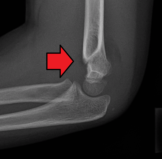
- Cartilaginous distal humerus and unossified capitellum make radiographs confusing
- Radius and ulna displaced together, almost always posteromedially
- Proximal radius stays aligned with the capitellum, which moves with the forearm
- Dislocation loses the radiocapitellar relationship and is rare at this age
- Lateral condyle fracture loses the relationship between capitellum and radius and ulna
- Ultrasound is quick, avoids anaesthesia and confirms the diagnosis in infants
- Arthrography under anaesthesia outlines the epiphysis and confirms reduction
- MRI when the diagnosis is still unclear
- Skeletal survey and child protection assessment when non-accidental injury is suspected
Management
- Undisplaced or minimally displaced fractures in an above elbow cast for about 3 weeks
- Displaced fractures treated with closed reduction, arthrography and percutaneous lateral or crossed pinning
- Pinning follows supracondylar fracture technique
- Late presentations beyond about a week left alone, as manipulation risks physeal damage
- These fractures remodel well
- Osteotomy later for residual deformity
Complications
- Cubitus varus is most common, from medial tilt and malunion of the epiphysis
- Avascular necrosis of the trochlea
- Growth disturbance
- Missed non-accidental injury, with risk of further harm to the child
Reviewed by Professor Phong Tran, Head of Orthopaedic Surgery, Western Health. Last updated 10 October 2026.