
Anatomy
Bone
- Organic phase mainly type I collagen, with proteoglycans, osteocalcin and osteonectin
- Hydroxyapatite gives compressive strength, collagen gives tensile strength
- Osteoblasts (mesenchymal origin) form osteoid
- Osteocytes sense load within lacunae linked by canaliculi
- Osteoclasts (haematopoietic origin) resorb bone in Howship lacunae
- Cortical bone forms osteons around Haversian canals
- Cancellous bone is trabecular with higher turnover
- Woven bone is immature and disorganised, lamellar bone mature and stress oriented
Articular cartilage
- No blood vessels, nerves or lymphatics, nourished by diffusion from synovial fluid
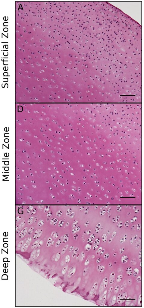
- Type II collagen and aggrecan, whose charged glycosaminoglycans attract water and resist compression
- Superficial zone collagen parallel to the surface, resisting shear
- Middle zone collagen oblique
- Deep zone collagen perpendicular, with the highest proteoglycan content
- Tidemark and calcified cartilage anchor to subchondral bone
- Fibrocartilage, mainly type I collagen
- Circumferential fibres resist hoop stress, radial tie fibres resist longitudinal splitting
- Peripheral portion vascular from the perimeniscal plexus, inner portion avascular
Tendon
- Type I collagen in fibrils, fibres and fascicles with aligned tenocytes
- Surrounded by paratenon or a synovial sheath
- Direct insertion through four zone fibrocartilage, or indirect via Sharpey fibres
Ligament
- More type III collagen and elastin than tendon, with less regular fibres
- Mechanoreceptors contribute to proprioception
Skeletal muscle
- Multinucleated fibres wrapped by endomysium, perimysium and epimysium
- Sarcomere between Z lines, with thin actin and thick myosin filaments
- Calcium from sarcoplasmic reticulum binds troponin, exposing actin binding sites
- Type I fibres slow twitch and oxidative for endurance
- Type II fibres fast twitch and glycolytic for power
Peripheral nerve
- Endoneurium surrounds axons, perineurium fascicles and epineurium the nerve
- Schwann cell myelin allows saltatory conduction between nodes of Ranvier
Pathology
- Tendon heals through inflammatory, proliferative and remodelling phases, never regaining full strength
- Ligaments fail at insertions in children and in midsubstance in adults
- Seddon classifies nerve injury as neurapraxia, axonotmesis and neurotmesis
- Wallerian degeneration occurs distally, with regeneration at about 1 mm per day
Reviewed by Professor Phong Tran, Head of Orthopaedic Surgery, Western Health. Last updated 10 October 2026.