
Definition
- Benign hyaline cartilage tumour on the bone surface beneath the periosteum
- Surface counterpart of enchondroma
Epidemiology
- Children and young adults, with slight male predominance
- Proximal humerus, proximal and distal femur, hands and feet
Pathology
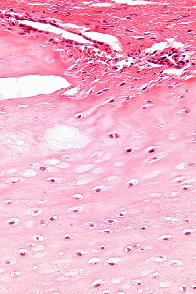
- Lobules of hyaline cartilage on the cortical surface
- Increased cellularity, binucleate cells and mild atypia are acceptable in this lesion
- No continuity with the medullary canal
- IDH1 or IDH2 mutations in a proportion
Clinical Features
- Slowly growing firm swelling
- Mild pain or tenderness
- Visible finger lump that may affect function
Investigations
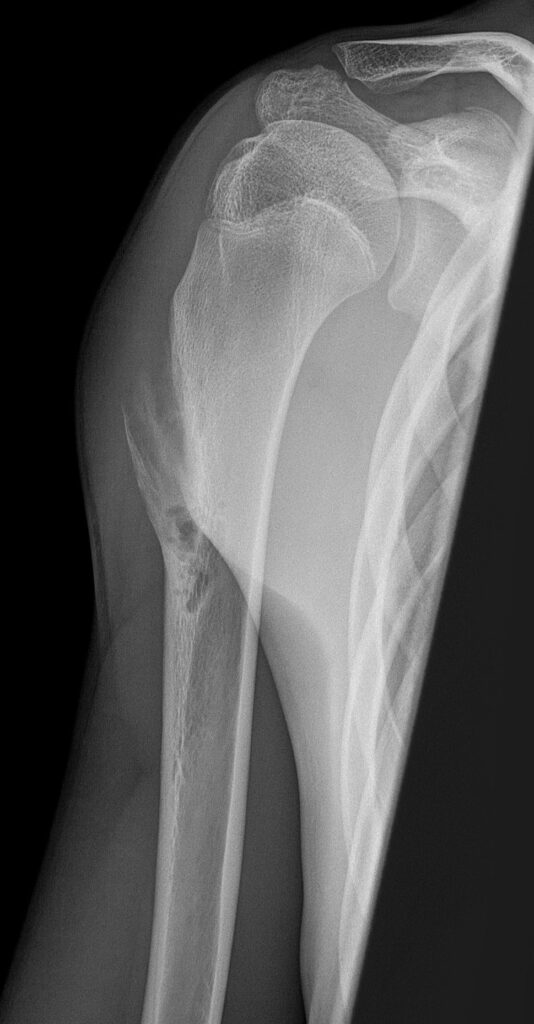
- Small surface lesion with saucerisation of the underlying cortex
- Sclerotic base with overhanging periosteal buttresses
- Rings and arcs calcification may be present
- CT and MRI confirm an intact medullary cavity
- MRI shows a lobulated high T2 lesion with peripheral and septal enhancement
- Most are under 3 cm
- Size over about 5 cm, cortical destruction, medullary invasion or soft tissue mass suggest chondrosarcoma
Differential Diagnosis
- Periosteal chondrosarcoma
- Periosteal osteosarcoma, larger with perpendicular spicules in tibial or femoral diaphysis
- Osteochondroma, with corticomedullary continuity
- Nora lesion (bizarre parosteal osteochondromatous proliferation) in the hands
- Soft tissue chondroma
Management
- Observe small asymptomatic lesions with typical imaging
- Marginal en bloc excision with underlying cortex for symptomatic or enlarging lesions
- Graft the defect if needed, fixation rarely required
- Curettage alone has higher recurrence
- Refer uncertain cases to a bone tumour unit before surgery
Prognosis
- Excellent, with uncommon recurrence after complete excision
- Malignant transformation is very rare
- Repeated recurrence or growth after maturity prompts reassessment for chondrosarcoma
Reviewed by Professor Phong Tran, Head of Orthopaedic Surgery, Western Health. Last updated 10 October 2026.