
Also known as
- hereditary multiple osteocartilaginous exostoses
- diaphyseal aclasis
Epidemiology
- Rare
Aetiology
- Autosomal dominant.
- Associated with mutations in EXT1 & EXT2, which leads to a maldistribution of Indian hedgehog in the extracellular matrix
- Usually associated with short stature
Clinical
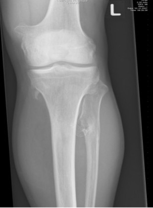
- Clinically multiple osteochondromas.
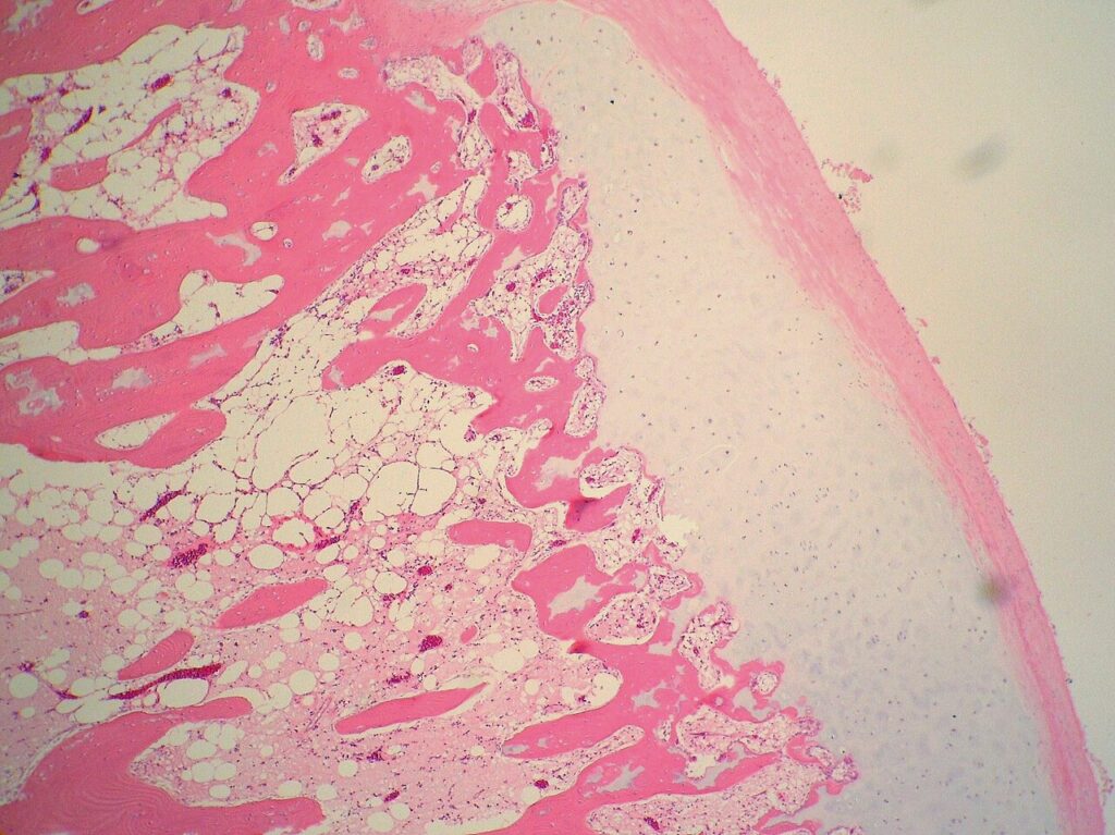
- Lesions are radiologically, grossly & microscopically similar to solitary osteochondromas
- A good sign is defects of tubulation of the long bones, particularly around the femoral neck
Malignant Transformation
- THERE IS A MUCH HIGHER RATE OF MALIGNANT TRANSFORMATION – AROUND 2-5%
- The presence of a thick, active cartilage cap on an exostosis in a skeletally mature individual alerts one to the possibility of malignant transformation
Reviewed by Professor Phong Tran, Head of Orthopaedic Surgery, Western Health. Last updated 10 October 2026.