
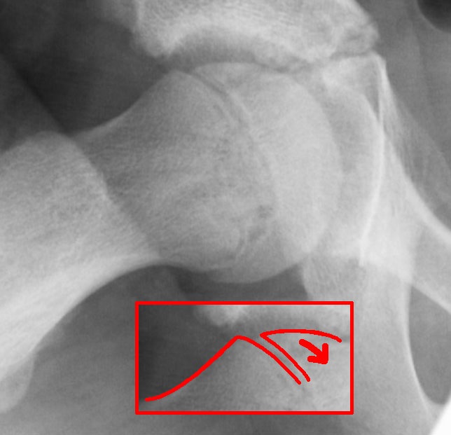
Epidemiology
- Incidence between 0.2-10/100 000
- M>F 60%:40%
- Mean age of diagnosis 13.5 in boys, 12 in girls
- Obesity can lead to earlier age of presentation
- 18-50% of cases can be bilateral
- Of the unilateral cases that go on to develop contralateral SUFE, second slip usually occurs within 18 months of the first
Aetiology
- Mechanical factors
- Obesity–> increased shear stress across the physis (obesity is associated with decreased femoral anteversion)
- Femoral Retroversion
- Increased physeal obliquity
Pathology
- Rapid longitudinal growth during puberty due to increased levels of growth hormone
- Rapid growth associated with increased chondrocyte proliferation and increased height of zone of hypertrophy. Increased height may contribute to decreased physeal strength
- Oestrogen reduces physeal height and increases physeal strength while testosterone reduces physeal strength–> May explain increased incidence in males
Natural History Without Treatment
- Unpredictable
- Risk of progression
- Risk of degenerative joint disease
Classification
Traditional (temporal) classification
| Type | Presentation | Examination |
|---|---|---|
| Pre-slip | Lower limb weakness, limp and exertional groin, hip or knee pain. AP pelvis and frog leg lateral may show disuse osteopenia | Reduced internal rotation and guarding |
| Acute (10 to 15%) | Under 3 weeks, usually too painful to weight bear. Most have 1 to 3 months of prodromal knee, hip or thigh pain or limp | External rotation deformity, shortening and painful restricted motion |
| Chronic (about 85%) | Groin or thigh pain with a limp, with exacerbations and remissions over months to years | Antalgic gait, loss of internal rotation, abduction and flexion |
| Acute on chronic | Acute worsening of a chronic slip | As for an acute slip |
Loder classification (physeal stability)
| Type | Definition | Osteonecrosis |
|---|---|---|
| Stable | Can weight bear, with or without crutches | Almost 0% |
| Unstable | Unable to weight bear, pain limits any attempt to move the limb | Up to 50% |
Original publication Loder RT, Richards BS, Shapiro PS, Reznick LR, Aronson DD. Acute slipped capital femoral epiphysis: the importance of physeal stability. J Bone Joint Surg Am. 1993;75(8):1134-40.
Treatment
- Stable SUFE
- In situ fixation
- Single Screw fixation- Most accepted treatment
- Advantage: Percutaneous placement with minimal soft tissue trauma. High success rate, low incidence of slip progression, osteonecrosis and chondrolysis
- Multiple screw/pin fixation
- Higher rate of unrecognised screw penetration compared to single screw
- Single Screw fixation- Most accepted treatment
- Bone graft Epiphysiodesis
- Avoid complications associated with internal pin fixation inc unrecognized pin penetration and damage to lateral epiphyseal vessels, although larger scar and increased blood loss and surgical time compared to in situ fixation
- Hip Spica Cast
- Immobilisation in bilateral hip spica avoids complications associated with surgical management. Risk of pressure sores, chondrolysis and further slip
- In situ fixation
- Unstable SUFE
- Risk of osteonecrosis higher than stable SUFE
- Consider urgent hip joint haematoma aspiration, closed reduction and single screw fixation
- Followed by 6-8/52 non-weightbearing
- Prophylactic fixation of contralateral side
- Risk of contralateral side becoming affected reported as 2335 times higher than the risk of the initial SUFE
Complications
- Osteonecrosis; increased with unstable SUFE, over-reduction, attempted reduction of stable SUFE, pin placement in the posterosuperior quadrant and cuneiform osteotomy
- Chondrolysis- usually secondary to unrecognised pin penetrations through the femoral head
Reference: Aronsson et al ‘Slipped Capital Femoral Epiphysis: Current Concepts’. J Am Acad Orthop Surg 2006; 14:666-679
Reviewed by Professor Phong Tran, Head of Orthopaedic Surgery, Western Health. Last updated 10 October 2026.