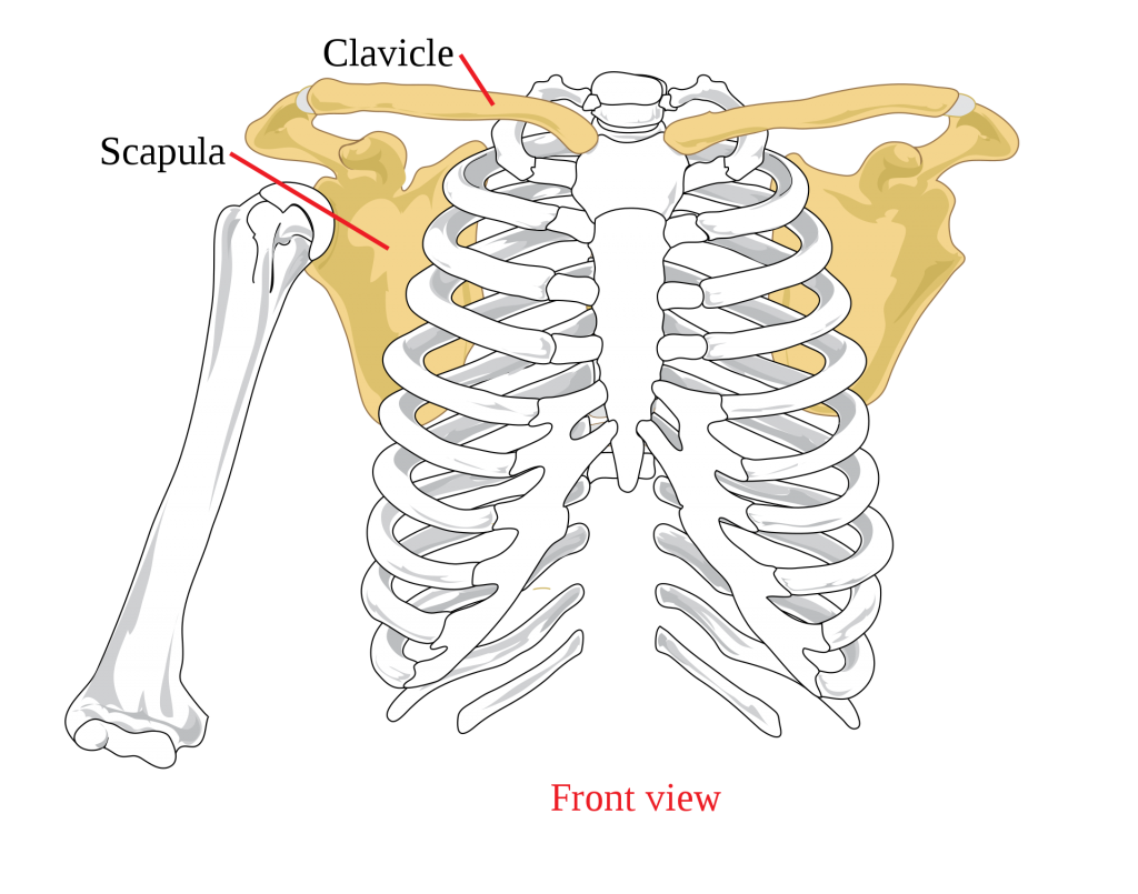
- Pectoral girdle connects the upper limb to the axial skeleton
- Consists of the clavicle and the scapula
- Two joints – sternoclavicular and acromioclavicular
- Strong coracoclavicular ligaments attach the clavicle to the scapula
Movements
- Movement of the glenohumeral joint is accompanied by movement of the clavicle and scapula
- Glenohumeral joint: flexion / extension, adduction / abduction, rotation
- Clavicle: moves in both horizontal and coronal planes, passive rotation around the sternoclavicular joint
Muscles
- Pectoralis Major
- Two heads, sternal (C7, C8, T1) and clavicular (C5, C6)
- Forms the anterior axillary wall
- Origin: Sternal half of clavicle, sternum to 7th rib, cartilages of true ribs (upper 6), aponeurosis of external abdominal oblique muscle
- Insertion: Lateral lip of the intertubercular sulcus of the humerus
- Primary Action: Flexes, adducts and medially rotates the arm
- Innervation: Medial and lateral pectoral nerves
- Pectoralis Minor
- Deep to pectoralis minor
- Delineates cords of the brachial plexus and parts of the axillary artery
- Origin: Outer surface of upper margin of ribs 3-5
- Insertion: Coracoid process of scapula
- Primary Action: Lowers lateral angle of scapula and protracts the scapula
- Innervation: Medial pectoral nerve
- Subclavius
- Origin: Costochondral junction of the 1st rib
- Insertion: Subclavian groove on the inferior surface of the clavicle
- Primary Action: Assists in stabilising the clavicle
- Innervation: Nerve to subclavius (C5, C6)
- Trapezius
- Assists shoulder abduction by rotating the scapula
- Origin: Superior nuchal line, external occipital protuberance, nuchal ligament, spinous processes of C7-T12
- Insertion: Lateral third of the clavicle, acromion, spine of scapula
- Primary Action: Elevate, retracts and rotates scapula, lower fibres depress scapula
- Innervation: Spinal part of the accessory nerve CNXI (C1-C5 or C6)
- Latissimus Dorsi
- Forms lower border of the posterior axillary fold
- Origin: Spinous processes of T7-L5, thoracolumbar fascia, iliac crest, and last three ribs
- Insertion: Floor of the intertubercular sulcus of humeus
- Primary Action: Extends, adducts, and medially rotates humerus
- Innervation: Thoracodorsal nerve (C6, C7, C8 – posterior cord of the brachial plexus)
- Rhomboid Major
- Origin: Spinous proccesses of T2 – T5
- Insertion: Medial border of scaphulabelow base of spine of scapula
- Primary Action: Retracts and rotates scapula to depress glenoid cavity
- Innervation: Dorsal scapular nerve
- Rhomboid Minor:
- Origin: Nuchal ligament, spines of C7 and T1 vertebraes
- Insertion: Medial border of scapula at spine of scapula
- Primary Action: Retracts and rotates scapula to depress glenoid cavity
- Innervation: Dorsal scapular nerve
- Levator Scapulae
- Origin: Posterior tubercles of transverse processes off C1-C4
- Insertion: Medial border of scapula from superior angle to spine
- Primary Action: Elevates scapula medially and inferiorly rotates glenoid fossa
- Innervation: Anterior rami of C3-C4 and dorsal scapular nerve
- Serratus Anterior
- Origin: Lateral surface of upper 8-9 ribs
- Insertion: Costal surface of medial border of scapula
- Primary Action: Protracts and rotates scapula
- Innervation: Long thoracic nerve
Joints
- Acromioclavicular Joint
- Sternoclavicular Joint
References
Last, R. and McMinn, R., 1994. Last’s Anatomy. 9th ed. Edinburgh: Churchill Livingstone.
Netter, F., 2018. Atlas Of Human Anatomy. 7th ed. Elsevier.
Author Contributions
Phoebe Walker